Diagnosis of Pulpal Pathology, General Updates

Overdiagnosis in Endo? Surely not!

I’ve noticed that the issue of overdiagnosis is really getting a lot of attention in the medical world. Overdiagnosis occurs when a person is diagnosed with a disease, but that disease, if left untreated would never have caused symptoms or death. This results in treatment being provided to people when they could have been left alone and never would have suffered. It is especially problematic when testing is involved that returns abnormalities that can be interpreted to indicate disease, but the test is inaccurate. It is also especially problematic when the treatment has significant consequences or side effects.

There was a conference held on the topic last year in the UK and I see this year’s conference is sold out. httpv://www.preventingoverdiagnosis.net/

I must admit that after reading a bit on the topic, there are a lot of aspects of overdiagnosis that apply to dentistry, as we are so focused on prevention of disease, and we do a lot of tests (pulp testing, bitewing and PA radiographs, probing of fissures) that can return both false positives and false negatives. Also, our treatments are invasive, costly and often irreversible.

This youtube video gives an easy to understand overview of testing: https://www.youtube.com/watch?v=U4_3fditnWg.

What are people’s thoughts on this? Do you think we are guilty?

Clinical Tips, Diagnosis of Pulpal Pathology, Endodontic Radiography

Perio-Endo? Get Some GP in it!

The second most common reason for extraction of root filled teeth is periodontal disease. So it’s important we identify periodontal disease when deciding if we should treat a tooth. Periodontal pockets around teeth can generally be due to three things:

1. Periodontal disease;

2. Vertical root fracture;

3. A sinus tract draining through the PDL.

Usually, a true periodontal pocket will be broad, whereas a pocket due to a root fracture or a draining sinus will be very narrow. We are more likely to find a true periodontal pocket in a patient who suffers periodontal disease in other teeth besides the one we are looking at.

When we do identify a periodontal pocket in a tooth that also has apical periodontitis, we want to know if the periodontal disease is separate to the apical disease. When these two are combined in a perio/endo lesion, the success rate in treating the teeth is lower than when we have separate perio and endo disease. If the pocket is narrow, then it’s important to differentiate between the draining sinus (which may be easy to treat) and the root fracture, which cannot be treated. I usually do this by raising a flap and inspecting the root with the microscope after staining with methylene blue.

Endo Perio lesion

GP placed into a sinus tract helps determine the true depth of the sinus tract.

In cases where I find a very deep, broad periodontal pocket which I think may be to the apex of the tooth, I place a GP point in the pocket to full length and take a radiograph. If the GP extends to the apex, then I know we have a fight on our hands to save the tooth.

The Endospot

A true perio/endo lesion makes this tooth a difficult prospect for successful treatment.

In the comments section, please leave your tips on identifying perio/endo lesions.

Diagnosis of Pulpal Pathology, Microbiology

How Strong Can A Pulp Be?


Way back in 1965 Kakaehashi et al(1) showed us that in the absence of microorganisms, even exposed dental pulps can survive quite well. For those of you who aren’t familiar with the research, gnotobiotic (or germ-free) rats had pulps exposed and the access cavities were left completely open. These rats lived in a sterile environment and ate sterile food. After the rats were sacrificed, histology showed that even though the pulp exposures were open and had food and debris packed into them, the pulps in these teeth remained vital and showed healing in the form of dentine bridging. Conversely, the rats who lived in normal environments showed bacterial contamination of their pulps leading to necrosis and paeriapical inflammation.

Vital Pulp Post Endodontic

This tooth had been asymptomatic for forty years, despite having no root filling present.

I recently treated a case where a single piece gold inlay and post needed to be removed as there was carious breakdown of the margins. The tooth was asymptomatic and had been so since the post and restoration was placed over forty years prior. There was no root filling to be seen .On removal of the post and restoration, I was a little surprised by what I found. Have a look at the video below…..

httpv://www.youtube.com/watch?v=1bgUq588xPc

The apical portion of the pulp remained vital after 40 years. This shows the importance that microorganisms play in pathology of the pulp. Other factors, especially heat from preparations (especially when not using water spray) and mechanical trauma can damage a pulp, but microorganisms play the MOST important role.

So, my recommendations when placing restorations are to use chlorhexidine or sodium hypochloride to clean cavities prior to restoration, use a good aseptic technique to exclude saliva (i.e. rubber dam) and use materials and techniques that seal well and prevent bacteria gaining access to deeper parts of restorations and thus the pulp. Oh, and don’t try putting a post into tooth without root filling it first. You’re not going to get as lucky as the dentist who did this one.

Endodontics, endospot

Final Obturation with post space left in tooth 21.


References:

1. The Effects of Surgical Exposures of Dental Pulps in Germ-Free and Conventional Laboratory Rats. KAKEHASHI S, STANLEY HR, FITZGERALD RJ. Oral Surg Oral Med Oral Pathol. 1965 Sep;20:340-9.

Diagnosis of Pulpal Pathology, Study Guides

Classifying Cracked Teeth. Operation Complex Part II

CRACKED TEETH: Classification

Cracked teeth can be one of the most complex, confusing and frustrating dental problems we face in every day practice. Its estimated in general practice that at least one patient per week presents with symptoms relating to a cracked or fractured tooth.  Accurate diagnosis and correct management are obviously the crucial steps of successful treatment, but we need to start by defining the various crack types to ensure we are all talking about the same thing at the same time.

It is important for us to have a clear understanding of what is meant by each definition as this allows discussion of the characteristics, prognosis and reliable treatment planning for each crack type. There are many classification systems out there for cracked teeth. Thankfully, the American Association of Endodontists (AAE) has developed a simple classification system for longitudinal tooth fractures based upon their location, direction, and extent. Not everyone out there agrees with this classification, but it’s the best we have and a good starting point when trying to manage.

Craze Lines
Craze lines occur only within enamel. They run parallel to enamel rods and terminate at the DEJ (Bodecker et al. 1951). Craze lines are present in most adult teeth. Various patterns of infraction lines can be seen depending on the direction and location of the impact to enamel, i.e. horizontal, vertical or diverging. Anterior teeth often exhibit vertical craze lines, involving the incisal edge or proximal corners. Posteriorly, craze lines usually cross the marginal ridges and extend along buccal and lingual surfaces.

Fractured Cusp
Fractured cusps occur most frequently in heavily restored teeth, where the marginal ridge is weakened and the affected cusp has insufficient support (Kahler 2008). A fractured cusp involves a complete or incomplete fracture initiated from the crown and extending subgingivally, usually directed both mesiodistally and buccolingually (Rivera & Walton 2008). The fracture usually crosses the marginal ridge, and also tracks down a buccal or lingual groove. It extends to the cervical third of the crown or root.

Cracked Tooth

Fractured Cusp

Cracked Tooth
A cracked tooth is an incomplete longitudinal fracture originating in the crown and extending apically. Often cited as being only in a mesio-distal direction (Rivera & Walton 2008), the literature also reports an significant number of bucco-lingual fracture planes (Seo et al. 2012). It may extend through either or both of the marginal ridges, through the proximal surfaces and onto the root surface. Occlusally, the crack is more centred and apical than a fractured cusp and therefore more likely to cause pulpal and periapical pathosis (Rivera & Walton 2008). A cracked tooth may progress to a split tooth.

Cracked Tooth

Cracked Tooth

Split Tooth
A split tooth is a complete fracture originating in the crown and extending subgingivally, directed most commonly mesiodistally through both marginal ridges and proximal surfaces (Seo et al. 2012). The split root area is often in the middle or apical third and tends towards the lingual. The more centred the crack is on the occlusion, the further apically the split extends. The segments are entirely separate and although it may occur suddenly it may be considered as a continuum from an incompletely cracked tooth (Rivera & Walton 2008).

Split Tooth

Split Tooth

Vertical Root Fracture
Vertical root fractures are complete or incomplete fractures initiated from the root (at any level), usually directed buccolingually (Rivera & Walton 2008). Most occur in endodontically-treated teeth although the literature reports occurrences in non-root filled teeth (Yang et al 1995). A VRF may progress coronally and/or apically from the point of origin in any part of the root.

Vertical Root Fracture

Vertical Root Fracture

Now we’re all on the same page, take a look at the next post in this series: Management of Cracked Teeth for an overview of recommended treatment strategies.

REFERENCES
Bodecker, CF, Gottlieb, B, Orban, B, Robinson, HB, Schour, I,  Sognnaes, RF 1951. Enamel lamellae. Oral Surg Oral Med Oral Pathol, vol. 4, 787-98.
Kahler, W 2008. The cracked tooth conundrum: terminology, classification, diagnosis, and management. American Journal of Dentistry, vol. 21, 275-82.
Rivera, EM,  Walton, RE 2008. Cracking the Cracked Tooth Code: Detection and Treatment of Various Longitudinal Tooth Fractures. Endodontics Colleagues for Excellence, Summer 2008.
Seo DG, Yi YA, Shin SJ, Park JW (2012) Analysis of factors associated with cracked teeth. Journal of Endodontics 38(3), 288-292.
Yang SF, Rivera EM, Walton RE (1995) Vertical root fracture in nonendodontically treated teeth. Journal of Endodontics 21(6), 337-339.

Aetiology of Apical Periodontitis, Clinical Tips, Diagnosis of Pulpal Pathology

Viral Pulpitis. This Would Have to Hurt…..


We usually blame bacteria for causing pulpitis but………. This patient complained of severe irreversible pulpitis symptoms for four days, which were only just starting to settle. She couldn’t isolate to either the upper second premolar or molar. Her dentist had extirpated the molar but symptoms hadn’t changed. Pulp testing showed the premolar was also responding negatively.

Take a look at the image below. This presentation is consistent with herpes zoster, commonly referred to as shingles. It is caused by the varicella zoster virus, also responsible for chicken pox. The trigeminal nerve is an unusual location for an outbreak (approximately 1-2% of cases), but when it occurs the virus affects a particular dermatome. If this dermatome also includes teeth, the pulp can also be affected. I can only imagine how painful this would be. Long term follow up is required as multiple teeth may lose vitality.

Notice how the viral outbreak is only affecting the greater palatine nerve distribution. If the nerve innervating the teeth are involved, this can lead to pulpitis and necrosis of the pulp tissues.

Diagnosing this initially may prove difficult, as the lesions on the mucosa don’t show up until a few days after symptoms appear, but some things to look out for include pulpitis symptoms from multiple teeth and a tingling or burning sensation in the distribution of the trigeminal nerve.

Pat Caldwell

Clinical Tips, Diagnosis of Pulpal Pathology

Pulp Testing – An Endodontist’s Best Friend

Back when I was working as an Endodontist with the Australian Navy, I often mentored newly graduated dentists. There was an interesting interaction between us that would often occur. The junior dentist would come into my surgery with a question about diagnosis. They would explain a set of symptoms and then show me a radiograph. I would then ask what the pulp test result was. The new grad would then slowly back out of the room in order to return to the patient and conduct the test.

I’m not sure why the pulp testing was left off the list, but I hope I hope that by the time the junior dentist had finished their posting and moved on to a new posting that pulp testing is one of the most important tools allowing us to to diagnose. Unfortunately, there is nothing certain when it comes to pulpal diagnosis, especially when we get to the grey area of reversible/irreversible pulpitis. If we really want to know what’s happening in the pulp and periapical area of a tooth, we need to extract it, section it, and look at it under a microscope. But that’s not practical…….

Pulp testing is not 100% accurate. If it was, then diagnosis would be much easier, but it often gives us essential information that will confirm a clinical diagnosis. When it comes to vital pulps, electric and cold testing gives a positive result 81-86% of the time (Petersson & Kiani-Anaraki, 1999). In a partially or completely necrotic tooth, these modalities will give a negative result on almost all occasions. This is useful infromation to have at hand.

Here is an example from when I was doing my post-graduate training:

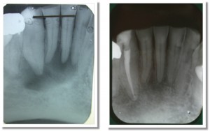
Images showing importance of pulp testing

Compare these radiographs. The image on the left is the pre-op and the image on the right is the two year review.

In the case above, we can see there is a definite lucency surrounding the lower right canine and central incisor. The lateral incisor may also be involved. Pulp testing with both cold and EPT revealed the canine was testing negative and the incisors were testing positive. As only the canine was testing negative, I only treated the canine. At the two year review, we can see that the lesion has healed and normal periradicular architecture has been re-formed around the vital incisors. If we just worked from the x-rays, we’d probably treat all three teeth, and subject the patient to unnecessary treatment. This is a pretty simple lesson.

Here are the keys to successful pulp testing:

1. Make sure the teeth are completely dry and isolated with cotton wool rolls;

2. Place the tip of the pulp tester on tooth structure, not restoration, and use a small (perhaps half of a) cotton pellet or endo sponge;

3. Start your test with a tooth that you expect will give a normal result and is not involved with the area in question. This will allow the patient to get a feel for what a normal response is;

4. Test with both cold and EPT. If the patient is experiencing symptoms such as a hyper-response to hot foods/liquids, then use hot as well.

In my surgery I have the pulp tester sitting right next to me, along with things like the apex locator and endo motor. We use it that often.

References:

Petersson KS, C. Kiani-Anaraki, M. Evaluation of the ability of thermal and electrical tests to register pulp vitality Endod Dent Traumatol 1999;15:127-131.

Diagnosis of Pulpal Pathology

The Endospot Simple Guide to Pulpal and Periapical Diagnosis Part 2

In this post we discuss a simple and practical method of classifying periapical pathology that can be used in practice on a daily basis.

Diagnosis of Apical Periodontitis

What’s Your Diagnosis?

Listen to the audio version here (5 mins, 50s) : Pulpal and Periapical Diagnosis

References referred to in the episode:

Abbot P. 2004. Classification, diagnosis and clinical manifestations of apical periodontitis. Endodontic Topics 2004, 8, 36–54

Andreasen FM. Transient apical breakdown and its relation to color and sensibility changes after luxation injuries to teeth. Endod Dent Traumatol. 1986 Feb;2(1):9-19.

Eliasson S, Halvarsson C, Ljungheimer C. Periapical condensing osteitis and endodontic treatment. Oral Surg Oral Med Oral Pathol. 1984 Feb;57(2):195-9.

Johnson W. 2002. Color atlas of Endodontics. W.B. Saunders, Philidelphia

Episode Transcipt:

Welcome to the Endospot episode 3. We’re now coming to you from Brisbane, Australia.

Well, the big news for me since the last episode is that I’ve moved home from Shanghai and have set up practice in Brisbane, Australia. It’s been an interesting process setting up a practice. There are lots of buying decisions to be made and I’ll probably talk about those in some upcoming blog posts so get on to the website at www.endospot.com and remember you can sign up to have the blog posts delivered directly to your email box.

Today we’re going to quickly go through the clinical classification system that I use for periapical pathology. If you’ve listened to the last episode, you’ll remember that I like to split up my diagnosis into pulpal and periapical diagnoses. If you haven’t listened to it, do so after this podcast is finished.

As with the pulpal diagnoses, there are quite a few different systems for classifying periapical conditions, and the one I like to use is similar to that published in Johnson’s clinical atlas of endodontics. I like it because it’s simple and easy to apply clinically, and will definitely help you arrive at a treatment plan.

The first diagnosis is the obvious one, “healthy periapex”. Usually, this will accompany a healthy pulp, there will be no symptoms of pain, no tenderness to percussion or palpation, and radiographically, the apex will appear completely normal. This is the diagnosis we like to make.

Diagnosis number two is “acute apical periodontitis”. This is an acute diagnosis, so by definition, there will be pain associated with this. And that means pain on percussion and possibly biting as well. Radiographically, the apex should appear normal or at most there might be some thickening of the PDL space. Importantly, you should realize that acute apical periodontitis will often occur along with pulpitis because when inflammation of the pulp occurs, we get an associated inflammation of the periapical tissues. But, the one to watch out for is where we have a normal pulp, and the periapical area is inflamed due to something like occlusal trauma or acute sinusitis. Obviously in this case performing root canal treatment isn’t going to lead to resolution of the symptoms.

The third diagnosis is ‘chronic apical periodontitis’. So the difference here is the word chronic. By definfition this is not a painful condition. This diagnosis is almost always accompanied by a necrotic pulp and the infection in the pulp is causing a periapical lesion which is picked up on a radiograph. In most cases, the pulp test will be negative, but occasionally, if the pulp is only partially necrotic, then you may still get a positive pulp test. The tooth may be slightly tender to percussion as well.

Our fourth diagnosis is ‘acute periapical abscess’. So this is an acute diagnosis so we have pain, the pulp will be necrotic so we’ll usually get a negative pulp test, there should be a lucency obvious radiographically. With a diagnosis of abcess the tooth will be very tender to percussion, often even with a gentle finger tap, and the tooth may be painful to bite on and may even be elevated in it’s socket. There can be localized swelling and lymph node enlargement as well.

Our fifth diagnosis is “chronic apical abscess”. Again because this is a chronic condition, there shouldn’t be symptoms of pain, and the key finding here is that of a sinus tract. The pulp will usually be completely necrotic, so expect a negative pulp test and a radiographic lucency. Because this diagnosis is due to the tooth being associated with an abscess, but the pus that is forming is being released through the sinus tract, these teeth are rarely symptomatic at all. You should be able to place a GP point into the sinus tract and take an x-ray to trace the sinus to a root tip.

The sixth and final diagnosis I want to talk about today is condensing osteitis. You don’t find a lot of information about this in the literature. It’s a diagnosis that you make radiographically by seeing an increased density of bone around an apex. Usually this will be associated with a pulp that is chronically inflamed but still vital, and the body has responded by increasing the bone density around the apex. I’ll put a reference for a case series in the show notes on the website.

So that’s it for periapical diagnosis. You need to be aware that twhat I’ve described is is a simple clinical diagnosis classification system designed to help you determine what treatment should be provided. There are always exceptions and you might find situations that don’t exactly fit for example when an abscess forms very quickly after pulpal necrosis but the periapical lesion doesn’t expand to a size that produces a lucency on a radiograph. Certainly the diagnoses we discussed tend to require a radiographic component, but it should be the clinical picture and symptoms that your patient is describing which heads you towards your diagnosis. There are also a bunch of conditions that can present as a lucency on an xray which are not endodontic in origin so again, your clinical history and tests should fit the radiographic appearance.

If you want a thorough review of the interaction of the periapical pathologies then go and grab Abbot’s article in Endodontic Topics, reference will be in the show notes. There are also some less common situations where other diagnoses are appropriate such as transient apical periodontitis and if you want to look that up I’ll put the reference for that in the notes too.

And in the meantime, remember you can go to the site and sign up to receive the endospot directly into your email box. That’s it for today. I’ll look forward to speaking to you next time.

Diagnosis of Pulpal Pathology, Endodontic Radiography, General Updates

Simple Guide to Pulpal and Periapical Diagnosis Part 1

In this post, we discuss a simple and practical method of classifying pulpal pathology that can be used in practice on a daily basis. For the new and updated Endospot Podcast, go to the Podcast Page.
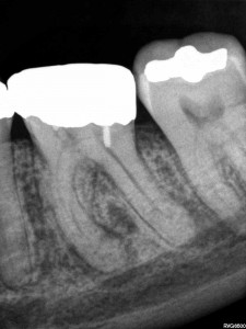
Endodontic diagnosis

How do you classify the pulp in this tooth?

Listen to the episode here (12 minutes):The Endospot Episode 2 | Simple Guide to Pulpal and Periapical Diagnosis Part 1

References Referred to in This Episode:

BARBAKOW F, CLEATON-JONES P, FRIEDMAN D. Endodontic treatment of teeth with periapical radiolucent areas in a general practice. Oral Surg Oral Med Oral Pathol 1981. 51, 552–559.

BENDER IB. 2000 Pulpal Pain Diagnosis. A Review. J Endod 2000. 26, 175-179

BENDER IB. 2000. Reversible and irreversible painful pulpitides: diagnosis and treatment. Aust Endod J 2000. 26, 10–14.

MILES TS. Dental pain: self-observations by a neurophysiologist. j Endod 1993. 19, 613-5.

SELTZER S, BENDER IB, ZIONTZ M. The dynamics of pulp inflammation: correlations between diagnostic data and actual histologic findings in the pulp. Oral Surg Oral Med Oral Pathol 1963. 16, 846-71 and 1963. 16, 969-77.

SIGURDSSON A. 2003. Pulpal Diagnosis. Endodontic Topics. 5, 12-25.

Transcript:

Welcome to the Endospot Episode 2.

My Name’s Pat Caldwell and today we’re coming to you from Shanghai, China. On our website we have the complete list of references discussed in this episode, so go to www.endospot.com and remember you can sign up online to receive the Endospot directly to your inbox.

In this episode, we’ll be discussing the classification of pulpal pathology. This will be the first in a series of three episodes looking at the issue of diagnosing pulpal and periapical pathology and in this episode we’ll go through a useful classification system which will help you deliver appropriate treatment to your patients. I’ve broken it up into three episodes because diagnosing and therefore treating appropriately is just so important. I think that often not enough time and thought goes into the initial diagnosis and that is when errors are made and our patients suffer. As endodontists we often see cases where an incorrect diagnosis has been made and treatment either withheld or provided when it wasn’t required and as professionals we owe our patient more than that.

When we look at the pulp and periapical classification systems, there are many out there, but in my opinion the simplest and most useful is the one proposed by Asgir Sigurdsson (Sigurdsson 2003). He published an excellent journal review article on this particular topic in everyone’s favourite journal, Endodontic Topics and that’s the classification system I’ll be discussing today. This classification is very much designed to give a clear indication of the treatment requirement according to the diagnosis, and when it comes down to it, that’s what we’re really looking to achieve out of our diagnosis.

Now when I’m faced with a new patient, whether they are presenting in pain or not, I will still conduct a complete history and examination and make both a pulpal and periapical diagnosis. I split the diagnosis into pulpal and periapical because it helps me have clear in my mind exactly what I’m dealing with, how we should proceed and what follow up’s required. It will also give us clues as to what problems we might run into, such as if the tooth will be difficult to anaesthetise, or if there’s likely to be a high level of post-operative pain. To be fair it will most often be the pulpal diagnosis that is driving your treatment decisions and that’s another reason to spend an episode on going through the various diagnoses.

Now if you think about it, the only way we can definitively diagnose the state of a pulp is to extract the tooth and slice it up and look at it through a microscope. So, our clinical diagnosis is always going to include an element of a guess and I can assure you that it will not always be straightforward. But I dIgress, we’ll get onto how to diagnose in a future episode so now let’s get back to the topic at hand. The classification system we are going to be using includes 4 different diagnoses for the dental pulp, and one of them is the healthy pulp. That’s the easiest one so we will start with that one.

A diagnosis of healthy pulp assumes that the pulp is vital and is not inflamed in any way. This tooth will respond normally to pulp testing, that is hot, cold and electric.  It will be asymptomatic and shouldn’t be tender to percussion or palpation unless there is an occlusion issue causing this. We use this classification sometimes when we need to do RCT on a tooth for prosthodontic reasons. So an example might be where there is not a lot of coronal tooth structure remaining in a vital tooth and the restorative dentist feels that an intracoronal restoration is required in order to provide retention for a core.

We’ll now move on to the more complex diagnoses, the ones indicating some sort of inflammatory response. The first of these is Reversible pulpitis. If we looked at this pulp under a microscope, we’d see a vital pulp with areas of localised inflammation. Most commonly this will be associated with a response to caries or possibly microbial leakage of a restoration. It could also be due to exposed dentine or bacterial ingress along a crack. All these things will lead to inflammation in the pulp. Now by definition, this inflamed pulp should heal when we remove the cause of the inflammation, so it’s important not to misdiagnose this pulp with an irreversible pulpitis and initiate RCT when it’s not needed.

A pulp with reversible pulpitis can present with quite a broad range of symptoms. Typically, the patient will describe pain with hot or cold, or with biting in the case of a crack. The pain might be mild, but can sometimes be severe, but probably the key to this diagnosis is that removal of the stimulus will lead to rapid relief of the pain. For example, drink something hot, tooth hurts, swallow the drink, tooth is fine. In general, there should also be no report of spontaneous  pain, and no tenderness to percussion or palpation. The tooth is likely to respond to a pulp test, but the inflammation in the pulp might mean that an exaggerated response is gained.  This in itself isn’t an indication of irreversible pulpitis, if the other indicators are that of reversible inflammation.

When you diagnoses reversible pulpitis and remove the stimulus by removing caries, or covering exposed dentine, it’s important that you review the patient and re-do all the examination procedures. Because if you think about, if you’ve diagnosed reversible pulpitis, say in case where there is a carious lesion and you treat it by removing the caries and placing a restoration, but the symptoms remain then by definition the pulp wasn’t reversibly inflamed.

Choosing between a diagnosis of reversible pulpitis, and it’s for more unpleasant alternative irreversible pulpitis is a critical decision, because the treatment options are very different. Where a pulp is irreversibly inflamed, this means that the inflammation is so severe that the pulp will not be able to heal. It will eventually necrose and become infected, leading to apical periodontitis. The treatment for these teeth is to undertake root canal therapy in order to remove the diseased pulp tissue and prevent infection.

As with reversible pulpitis we see a wide range of presentations. Commonly there is an exaggerated response to hot and cold, but the key here is that this response lingers for some time. It’s hard to say exactly how long it has to linger to be considered irreversible, so here I’ll have to take some licence and say that a pain that lingers for a number of minutes after stimulus is not a healthy pulp. There is a good biological explanation for this lingering pain but I’ll go through that with you in the diagnosis episode. Sigurdsson tells us to be careful when interpreting this symptom as if you put an intense stimulus even on a healthy pulp the resulting pain will linger somewhat.

Another indicator that we’re dealing with an irreversibly inflamed pulp is where the pain has been severe, and the longer it has been present, the more likely it is to be irreversible in nature (Bender 2000). When pulp testing this tooth, there will often be an exaggerated response and the dull lingering pain experienced before will be induced by the procedure. Tenderness to percussion is often also present. The level of inflammation in these teeth will often lead to neurogenic pain and nerve sprouting which results in inflammation in the periapical tissues. This can occur well before the pulp starts to die and therefore even in a vital, inflamed pulp, tenderness to percussion is often present.

Probably the biggest indicator of an irreversibly inflamed pulp is spontaneous pain. So the patient will report moderate to severe pain that suddenly occurs, and often remains as a dull lingering pain for minutes or hours. They might even report being woken by the pain. I often find that patients with irreversible pulpitis have resorted to over the counter painkillers such as ibuprofen and report that these drugs will help relieve the pain until they wear off of course.
There is an excellent article published by Timothy Miles, who is a neurophysiologist who had previously trained as a dentist (Miles 1993). In the article he explains in detail his not only the physiological response he personally experienced due to a dying pulp, but also the emotional response. It has a grand total of 6 references which it probably could have done without and yet was published in the Journal of Endodontics. It’s probably essential reading for any dentist who has never experienced toothache, and I’ll put the reference in the show notes. If you ever sit the examinations for the Royal Australasian College of Dental Surgeons, then I believe he still lectures on the primary orientation course, and you can have the pleasure of hearing the story in person.

Our final diagnosis is pulpal necrosis. This covers both partial and complete necrosis of the pulp. Consider the progression from reversible pulpitis to irreversible pulpitis and on to pulpal necrosis. At some point the inflammation builds to a level where the vital tissue dies. This necrosis over time then spreads throughout the whole pulp space. There isn’t a lot of information on exactly how quickly the process occurs and I imagine that there is a great variation in the length of that process, but when it comes down to it, some form of Root canal therapy is required.

Most of the time when a pulp is necrotic, it will also be infected. There are a few situations where a pulp can necrose and remain uninfected, mostly after physical trauma to a tooth, but on the whole, a dead pulp lacks a blood supply and therefore lacks the ability to protect itself against microorganisms. These bugs will get into the pulp space through cracks or infected dentine, or possible exposed dentinal tubules and rapidly take over the necrotic space.

When dealing with a patient presenting in pain, there is a strong correlation between the following factors and pulpal necrosis. First is a history of moderate to severe pain. Second is tenderness to percussion, third is a history of spontaneous pain and fourth is a negative pulp test. Seltzer and Bender conducted some of the most useful research we have on this topic way back in 1963 (Seltzer et al. 1963). Basically, they examined patients who presented with pain and then the tooth was extracted and examined histologically. There is a nice summary of their findings in a paper published by IB Bender in 2000 in the JOE which you should read if you are a postgraduate student (Bender 2000).

It’s important to note here though that very often the progression from vital to necrotic pulp is painless, or the level of pain is minimal and no help is sought for the problem. Often patients are completely unaware that they have a chronic infection in their jaw. Two studies reported this happening in 26-60% of cases. (BARBAKOW ET AL. 1981, BENDER 200) In my experience these teeth may be completely asymptomatic. They can be slightly tender to percussion but often don’t even present with this. Usually, they are identified due to a lucency on a PA or OPG Xray. The key for these though is that they will not respond to a pulp test and can be entered without local anaesthetic.

OK, I think that’s enough for this episode. Next episode we’ll be discussing the various periapical diagnoses that we can make. In the meantime, I recommend you have a look at the references, and next time and every time you work on a tooth, have a think about what diagnosis you have for the pulp. If you’re placing large restoration or crown think about whether you’re completely confident of your diagnosis before doing the work.

Also, if you’ve got a comment, or you disagree with something that was said here, please keep it nice, but I’d love you to go to the blog and leave a comment.