General Updates, Study Guides

Are Most Published Research Findings False?

Slimey DelightAlong with some other enlightening speakers, I just had the pleasure of listening to a full day of lecture material by Dr John Ioannidis. Dr Ioannidis is a leader in the field of Epidemiology and Research Statistics. In 2005 he authored an article entitled, “Why Most Published Research Findings are False”.

I’ve read his articles before, but having the man himself run through each of the areas covered in the article was a real eye opener. I don’t want to be critical of all the dedicated and hard-working researchers out there. We owe them a debt of gratitude. But, it would seem that the publishing of research findings is a an area where accidental (or non-accidental) misleading statement, conclusions and assumptions are often made.

The biggest issue we have is the pursuit of findings that are often based on research with a formal statistical significance, commonly with a p value less than 0.05. Dr Ioannidis shows that these findings can often be incorrect. He should know. He’s been involved in large scale studies which made findings that were later refuted and shown to be (more than likely) incorrect.

As practitioners, we are called upon ever day to make decisions based on the published research, so it is important that we have a good understanding of what we are reading. I recommend reading the article by Dr Ioannidis and taking note of the corollaries he mentions. It is this sort of introspection which makes us better practitioners.

Please, read his article. In fact, don’t read it, study it, and get interested in what all those statistics actually mean.

John Baez does a nice job of simplifying the corollaries, and there is some good discussion to follow it on his blog.

The article is free here: http://www.plosmedicine.org/article/info:doi/10.1371/journal.pmed.0020124

Diagnosis of Pulpal Pathology, Study Guides

Classifying Cracked Teeth. Operation Complex Part II

CRACKED TEETH: Classification

Cracked teeth can be one of the most complex, confusing and frustrating dental problems we face in every day practice. Its estimated in general practice that at least one patient per week presents with symptoms relating to a cracked or fractured tooth.  Accurate diagnosis and correct management are obviously the crucial steps of successful treatment, but we need to start by defining the various crack types to ensure we are all talking about the same thing at the same time.

It is important for us to have a clear understanding of what is meant by each definition as this allows discussion of the characteristics, prognosis and reliable treatment planning for each crack type. There are many classification systems out there for cracked teeth. Thankfully, the American Association of Endodontists (AAE) has developed a simple classification system for longitudinal tooth fractures based upon their location, direction, and extent. Not everyone out there agrees with this classification, but it’s the best we have and a good starting point when trying to manage.

Craze Lines
Craze lines occur only within enamel. They run parallel to enamel rods and terminate at the DEJ (Bodecker et al. 1951). Craze lines are present in most adult teeth. Various patterns of infraction lines can be seen depending on the direction and location of the impact to enamel, i.e. horizontal, vertical or diverging. Anterior teeth often exhibit vertical craze lines, involving the incisal edge or proximal corners. Posteriorly, craze lines usually cross the marginal ridges and extend along buccal and lingual surfaces.

Fractured Cusp
Fractured cusps occur most frequently in heavily restored teeth, where the marginal ridge is weakened and the affected cusp has insufficient support (Kahler 2008). A fractured cusp involves a complete or incomplete fracture initiated from the crown and extending subgingivally, usually directed both mesiodistally and buccolingually (Rivera & Walton 2008). The fracture usually crosses the marginal ridge, and also tracks down a buccal or lingual groove. It extends to the cervical third of the crown or root.

Cracked Tooth

Fractured Cusp

Cracked Tooth
A cracked tooth is an incomplete longitudinal fracture originating in the crown and extending apically. Often cited as being only in a mesio-distal direction (Rivera & Walton 2008), the literature also reports an significant number of bucco-lingual fracture planes (Seo et al. 2012). It may extend through either or both of the marginal ridges, through the proximal surfaces and onto the root surface. Occlusally, the crack is more centred and apical than a fractured cusp and therefore more likely to cause pulpal and periapical pathosis (Rivera & Walton 2008). A cracked tooth may progress to a split tooth.

Cracked Tooth

Cracked Tooth

Split Tooth
A split tooth is a complete fracture originating in the crown and extending subgingivally, directed most commonly mesiodistally through both marginal ridges and proximal surfaces (Seo et al. 2012). The split root area is often in the middle or apical third and tends towards the lingual. The more centred the crack is on the occlusion, the further apically the split extends. The segments are entirely separate and although it may occur suddenly it may be considered as a continuum from an incompletely cracked tooth (Rivera & Walton 2008).

Split Tooth

Split Tooth

Vertical Root Fracture
Vertical root fractures are complete or incomplete fractures initiated from the root (at any level), usually directed buccolingually (Rivera & Walton 2008). Most occur in endodontically-treated teeth although the literature reports occurrences in non-root filled teeth (Yang et al 1995). A VRF may progress coronally and/or apically from the point of origin in any part of the root.

Vertical Root Fracture

Vertical Root Fracture

Now we’re all on the same page, take a look at the next post in this series: Management of Cracked Teeth for an overview of recommended treatment strategies.

REFERENCES
Bodecker, CF, Gottlieb, B, Orban, B, Robinson, HB, Schour, I,  Sognnaes, RF 1951. Enamel lamellae. Oral Surg Oral Med Oral Pathol, vol. 4, 787-98.
Kahler, W 2008. The cracked tooth conundrum: terminology, classification, diagnosis, and management. American Journal of Dentistry, vol. 21, 275-82.
Rivera, EM,  Walton, RE 2008. Cracking the Cracked Tooth Code: Detection and Treatment of Various Longitudinal Tooth Fractures. Endodontics Colleagues for Excellence, Summer 2008.
Seo DG, Yi YA, Shin SJ, Park JW (2012) Analysis of factors associated with cracked teeth. Journal of Endodontics 38(3), 288-292.
Yang SF, Rivera EM, Walton RE (1995) Vertical root fracture in nonendodontically treated teeth. Journal of Endodontics 21(6), 337-339.

Clinical Tips, General Updates, Study Guides

An Endodontic Masterclass

This is just a short post to let you know that I will be running a hands-on course for general dentists on October 12 in Brisbane. I wanted to let Endospot followers have the first opportunity to sign up. It should be a great day with a nice mix of useful information, practical advice and hands-on experience.

You can download the course flyer here or sign up here.

I hope to see you there.

Pat Caldwell

Aetiology of Apical Periodontitis, Microbiology, Study Guides

The Lazy Man’s Guide to Persistent Apical Periodontitis

Poorly Prepared, Obturated and Missed Canals All Contribute to Persistent Apical Periodontitis

Persistent Apical Periodontitis (AP) refers to AP that is associated with a tooth that has had root canal therapy (RCT).  As with primary AP, bacteria are the most common cause of the inflammatory response (Sudqvist et al. 1998). Previously there has been a large body of evidence that persistent infections are commonly composed of a single species, however recent evidence points to the presence of a mixed biofilm (Siqueira et al. 2009a, Chavez de Paz 2007).”]

There are also non microbial causes of AP, including foreign body reactions, cystic formation, endogenous cholesterol crystals and scar formation. These will be discussed later.

The microbes that cause persistent AP are more commonly located intraradicularly (inside the root). Occasionally, these microbes will also be located extraradicularly. We’ll discuss the far more common intraradicular microbes first.

Intraradicular Microbes

The key study for referencing the presence of microbes within the root in cases of persistent AP is Nair et al. 1990a. When considering the cause of the persistent infection, consider that the microbes were either present prior to RCT being initiated (primary infection ) or they entered during or after treatment (secondary infection) (Siquiera 2008).  When thinking about those microbes that have survived from the primary infection, consider how they might have achieved this. They may have been resistant to the chemicals used in the disinfection process (e. feacalis for example has some mechanisms to survive calcium hydroxide), or they may have been located in a portion of the canal that was not instruments nor cleaned via chemical means.

When considering the secondary infection, these microbes may have gained access to the canal during treatment or after treatment. Think about this also. They may have been carried into the canal on a contaminated instrument or perhaps a leaking rubber dam may have allowed saliva to contaminate the root canal. Alternatively, a poorly placed temporary restoration may have allowed leakage into the root canal system in between visits. If caries have not been completely removed, or a previous restoration which is subject to microleakage is left in place, then this can also be a source of secondary infection. Alternatively, these microbes may have entered a previously clean  root canal system after the completion of RCT. This could be due to a leaking restoration, or through caries or a crack in the tooth. It’s important to understand the microbial nature of AP, and to have this foremost in our minds when undertaking treatment.

Which microbes are present in secondary in persistent AP?

When we look at the composition of the infection in AP, we find a significantly different microflora than that found in primary infections (Figdor et al. 2007, Molander et al. 1998). Generally in persistent AP, there are only 1-5 species.  These are predominantly gram-positive and there is an equal amount of obligate and facultative anaerobes. (Figdor et al. 2007, Sundqvist et al. 1998, Siqueira et al. 2009b). Due to the fact that obligate anaerobes are easier to kill, it may be that facultative anaerobes are more likely to persist within the root canal system after treatment.

E. Faecalis & c.albicans

Enterococcus faecalis is an opportunist pathogen which is implicated in many general surgery post-operative infections.  It has been identified as an opportunistic pathogen in persistent AP in a number of studies (Sundqvist et al. 1998, Sundqvist et al. 2003, Molander et al. 1998). This particular microbe has been studies extensively. It possesses a “proton pump” on its cell membrane which allows it to regulate its internal pH. This means that it is resistant to calcium hydroxide and this may be one of the ways that it survives and becomes implicated in persistent infections. It is also able to survive by itself and without nutrition for long periods of time. It is rarely found in untreated canals. Candida albicans (a fungus) is also found more commonly in persistent infections than in primary infections. (Sundqvist et al. 1998, Nair et al. 1990a, Waltimo et al. 2004)

Extraradicular Infections

Occasionally, we may find a situation where microbes establish themselves outside the root canal system. The microbes may establish themselves on the external root surface in a biofilm, in association with infected dentine chips that have been displaced into the periapical region, or within a periapical cyst (Abbott 2004, Nair 2008). These microbes must be able to withstand the body’s attempts to kill them and it is likely that biofilm formation allows this (Noiri et al. 2002). Similarly in the periapical cyst situation, it is the cyst itself that protects the microbe from the immune response.

In particular, two microbes have been implicated in extraradicular infections. These are Actonomyces spp. And Proprionibacterium proprionicus (Siqueira 2002). These microbes are able to form cohesive colonies within an extracellular matrix. This helps them to avoid phagocytosis, and so continue to survive and invoke the immune response.

Non-microbial causes of AP – Cysts, Foreign Body Reactions and Cholesterol Crystals

In some cases, AP may not be “maintained” by microorganisms. I say “maintained”, because often the AP is initially caused by microbes, and after endodontic treatment, one of the following factors takes over, maintaining the immune response and thus, AP.

Periapical cysts are an interesting topic. There are a range of studies that attempt to measure the incidence of periapical cysts in examined PA lesions. In simple terms, the lesion is biopsied and then examined under a microscope. If an epithelium lined sack is found, then the lesion is designated a cyst. But…… In 1980 Simon published a paper which included serial sectioning of periapical lesions (Simon, 1980). What he found was that some lesions that appeared as cysts on one section, appeared differently on other sections. Thus, it was deemed that the majority of studies (which don’t use serial sectioning) relating to the prevalence of cysts were subject to error. If you just take a random slice, the effect in two dimensions may be that of a cyst, when in reality the full three dimensional structure of the cyst does not exist. Nair repeated this study 16 years later and confirmed Simon’s findings (Nair, 1996).

Nair studied far more lesions than Simon, and found that 15% could be classified as cysts (including both true and pocket varieties). This is probably the best figure to be quoting. Other studies report figures from 5-55%, but they failed to use serial sections. It is also important to realize that a great number a large proportion of abcesses and granulomas will also contain epithelium. In Nair’s study, 52% of the lesions were epithelialised, but only 15% were cysts.  It is likely that the inflammatory process results in the proliferation of this epithelium, and over time, the epithelium develops into a cyst.

Through both of these studies, Simon and Nair found two distinct types of cysts. Simon called them true cysts, those with a complete epithelial lining, and bay cysts, those whereby the lining is attached to the root surface and the contents of the root canal contiguous with the contents of the cyst. Nair referred to these as true cysts and pocket cysts (equivalent to Simon’s Bay cyst).

Nair contends that these two types of cysts are quite different (Nair 2008). He feels that the true cyst is self sustaining, and will remain independent of efforts to remove the microorganisms from the root canal system. The pocket cyst, on the other hand is being sustained by the microbes within the canal system. Removal of the microbes which are maintaining the inflammatory response may allow the pocket cyst to heal. In reality, it will be very difficult to prove or disprove this theory, but one could say that it makes sense.

Foreign body reactions

When exogenous materials are located in the periapical region, they can induce and maintain an inflammatory response which may be asymptomatic, but will be seen as a radiolucency. Materials may be GP, amalgam, sealants, Calcium hydroxide or cellulose fibres such as those contained in paper points (Nair et al. 1990b).

In practice these lesions are rarely seen but have been reported in the literature, so it is important to understand that this mechanism for the maintenance of AP does exist. It also reminds us to be careful when using paper points not to extend them into the periapical areas, as human cells cannot degrade cellulose and leaving fibres behind may result in a foreign body reaction.

Gutta percha may also induce a foreign body reaction, especially in fine particles (Sjögren et al. 1995). Overextended GP may, as a result cause delayed healing of periapical tissues.

Cholesterol Crystals

Cholesterol crystals are also seen in AP, and are probably released by disintegrating erythrocytes, lymphocytes, macrophages, plasma cells and from circulating plasma lipids (Nair 1999). These collections of cholesterol are referred to as cholesterol clefts and induce a reaction similar to a foreign body reaction as the macrophages and giant cells are unable to remove the cholesterol. Again, this may result in a non-healing lesion, despite well completed endodontic treatment.

The Endospot Easy Study Guide to Persistent AP

  1. Persistent AP is most commonly caused by microbes remaining within the root canal system (sundqvist et al. 1998)
  2. It appears that a mixed biofilm may be responsible, contrary to the previous belief that usually only one microbe was responsible.  (Chavez de Paz 2007)
  3. The microbes are either (Siquiera 2008):
    1. Primary – remained within the canal from the initial infection
    2. Secondary – entered during or after treatment
  4. Significantly different flora to primary AP (Molander et al. 1998)
    1. 1-5 species per canal
    2. predominantly gram positive
    3. equal number of obligate and facultative anaerobes
  5. E. faecalis – opportunist pathogen which has been identified more commonly in persistent AP (Sundqvist 1998)
    1. Posesses a “proton pump” which allows it to survive in high pH (i.e. can survive calcium hydroxide)
    2. Can survive in mono-infection
    3. can survive long periods of low/no nutrition
  6. Candida Albicans also found more commonly in persistent infections than in primary (Waltimo et al. 2004)
  7. Exraradicular infections can occur in biofilm on the root tip (Noiri 2002), or in the PA area itself (Siquiera 2002)
    1. Proprionibactrium proprionicus and Acinomyces species are able to from adhesive colonies in an extracellular matrix in the PA tissues
  8. Non-microbial causes of AP are:
    1. Periapical cysts – 15% of lesions (Nair 1996)
      1. Serial sectioning indicates two types – true cysts and pocket cysts
    2. Foreign Body Reactions
    3. Cholesterol Clefts

References:

ABBOTT, P. V. 2004. Classification, diagnosis and clinical manifestations of apical periodontitis. Endodontic Topics, 30-54.

CHAVEZ DE PAZ, L. E. 2007. Redefining the persistent infection in root canals: possible role of biofilm communities. J Endod, 33, 652-62.

FIGDOR, D. & SUNDQVIST, G. 2007. A big role for the very small–Understanding the endodontic microbial flora. Australian Dental Journal, 52, 38.

MOLANDER, A., REIT, C., DAHLEN, G. & KVIST, T. 1998. Microbiological status of root-filled teeth with apical periodontitis. Int Endod J, 31, 1-7.

NAIR, P. N., SJÖGREN, U., KREY, G., KAHNBERG, K. E. & SUNDQVIST, G. 1990a. Intraradicular bacteria and fungi in rootfilled, asymptomatic human teeth with therapy-resistant periapical lesions: a long-term light and electron microscopic follow-up study. J Endod, 16, 580-8.

NAIR, P. N., SJÖGREN, U., KREY, G. & SUNDQVIST, G. 1990b. Therapy-resistant foreign body giant cell granuloma at the periapex of a root-filled human tooth. J Endod, 16, 589-95.

NAIR, P. N. 1996. Types and incidence of human periapical lesions obtained with extracted teeth. Oral Surg Oral Med Oral Pathol Oral Radiol Endod, 81, 93-102.

NAIR, P. N. 1999. Cholesterol as an aetiological agent in endodontic failures–a review. Aust Endod J, 25, 19-26.

NAIR, P. N. 2008. Pathobiology of Apical Periodontitis. Essential Endodontology. 2nd ed. Oxford: Blackwell Munksgaard.

NOIRI, Y., EHARA, A., KAWAHARA, T., TAKEMURA, N. & EBISU, S. 2002. Participation of bacterial biofilms in refractory and chronic periapical periodontitis. J Endod, 28, 679-83.

SIMON, J. H. S. 1980. Incidence of periapical cysts in relation to root canal. J Endod, 6, 845-8.

SIQUEIRA, J. F. 2002. Endodontic infections: concepts, paradigms, and perspectives. Oral Surg Oral Med Oral Pathol Oral Radiol Endod, 94, 281-93.

SIQUEIRA, J. 2008. Microbiology of Apical Periodontitis. Essential Endodontology. Oxford: Blackwell Munksgaard.

SIQUEIRA, J. F., JR. & ROCAS, I. N. 2009a. Community as the unit of pathogenicity: an emerging concept as to the microbial pathogenesis of apical periodontitis. Oral Surg Oral Med Oral Pathol Oral Radiol Endod, 107, 870-8.

SIQUEIRA, J. F. & RÔÇAS, I. N. 2009b. Diversity of endodontic microbiota revisited. Journal of Dental Research, 88, 969-81.

SJÖGREN, U., SUNDQVIST, G. & NAIR, P. N. 1995. Tissue reaction to gutta-percha particles of various sizes when implanted subcutaneously in guinea pigs. European Journal of Oral Sciences, 103, 313-21.

SUNDQVIST, G. & FIGDOR, D. 2003. Life as an endodontic pathogen. Endodontic Topics, 6, 3-28.

SUNDQVIST, G., FIGDOR, D., PERSSON, S. & SJÖGREN, U. 1998. Microbiologic analysis of teeth with failed endodontic treatment and the outcome of conservative re-treatment. Oral Surg Oral Med Oral Pathol Oral Radiol Endod, 85, 86-93.

WALTIMO, T. M. T., HAAPASALO, M., ZEHNDER, M. & MEYER, J. 2004. Clinical aspects related to endodontic yeast infections. Endodontic Topics, 66-78.

Aetiology of Apical Periodontitis, Microbiology, Study Guides

The Lazy Man’s Guide to the Microbial Causes of Primary Apical Periodontitis

R1This is a dry topic, so we’ll try to cover it as simply as possible. If you just want the outline, check out the study guide at the end of the blog post.

So Microorganisms cause Apical Periodontitis. How do they get into the tooth?

Primary apical periodontitis (AP) occurs when microorganisms (let’s call them bugs) enter the pulp chamber and colonise the pulp tissue. The bugs enter the pulp chamber through a variety of routes such as carious lesions, cracks, traumatic exposure of the pulp and due to dental procedures such as cavity preparation (Nair 1997). As mentioned in the previous post on the aetiology of AP, there is plenty of evidence of bugs being found in teeth that have apparently intact crowns (Bergenholtz 1974). These bugs gain access through accessory canals, exposed dentinal tubules, or microcracks adjacent to the gingival crevice or deep periodontal pockets (Nair 1997). The important message here is that we don’t require an obvious,visible path of entry to the pulp chamber. Bugs can enter through tiny, microscopic spaces such as dentinal tubules.

Continue reading

Aetiology of Apical Periodontitis, Study Guides

The Lazy Man’s Guide to the Aetiology of Apical Periodontitis

Lateral Incisor with Apical Periodontitis

What's the Root of This Problem?

Generally, apical periodontitis (AP) is a result of microorganisms entering the root canal system. Of course there are other causes of inflammation in the apical area, such as trauma, traumatic occlusion and foreign body reactions, but if we’re talking about common AP, then “bugs” are the cause (Abbott 2004, Harn et al. 2001).

The classic study on this topic was Kakehashi et al (1965), and this should be the starting point for any essay or exam answer. They took gnotobiotic rats (bacteria free rats) and conventional rats (with bacteria) and exposed the pulps in some teeth. In this study, the germ free rats had food, and in one case a hair forced into their long term exposed pulps, but necrosis did not occur! There was even dentine bridge formation in some cases. Conversely, all the conventional rats developed pulpal necrosis and apical periodontitis. It seems that without bacteria, rat pulps are very hardy indeed.

Sundqvist (1976) was the first to anaerobically culture and taxonomically analyse the constituent bacteria in teeth with necrotic pulps. He concluded that: AP is associated with the presence of bacteria in the root canal and that infected teeth harboured a mixture of bacteria that were predominantly obligate anaerobes (Sundqvist 1976). The significance of obligate anaerobes in endodontic infections are confirmed by a number of independent studies (Bergenholtz 1974, Wittgow et al. 1975, Kantz et al. 1974).

A study by Möller et al. (1981) compared the apical response of inducing pulp necrosis both aseptically and after contamination with indigenous oral flora in monkeys. Basically, they aseptically opened monkey pulps and used a Hedstrom file to mince the pulp. They then closed the pulp chamber so that the necrotic, uninfected tissue was inside, or they infected the tissue with plaque form the monkey’s teeth. In the teeth which contained sterile necrotic tissue, no AP formed. Those with bacteria were associated with AP. At this stage there was still a commonly held belief that necrotic tissue and stagnant tissue contributed to AP formation, but this study helps dispel this theory. In addition, this study showed that there was a variation in the ability of the various bacteria to survive and establish themselves within the root canal environment, and we start to see the concept of bacteria within root canals living in communities, rather than just as one individual strain.

There has been a significant shift away from the concept of one ‘causative pathogen’ and significant evidence exists that specificity seems to be related to the community level as certain species compositions are specifically associated with some forms of apical periodontitis (Siqueira et al. 2009a, Sakamoto et al. 2006). There is a very high level of inter-individual variation of bacterial community profiles, such that 44% of recovered taxa were isolated or detected in only one study (Siqueira et al. 2009b). Microbial ecology differs significantly between different disease forms such as chronic apical periodontitis versus acute apical abscess, suggesting the existence of a pattern associated with each one (Sakamoto et al. 2006, Siqueira et al. 2004). In addition, different magnitudes
of disease, based on intensity of signs and symptoms, may be related to the species composition of the community (Siqueira et al. 2009a).

Siqueira (2002) differentiates the types of endodontic infections into primary, secondary or persistent root canal infections.  Primary infections are those that occur within a root canal system prior to any treatment being applied. A Secondary infection occurs when a root canal (RC) had been treated, and new bacteria enter the RC system.  This can happen when a filling is lost, or micorleakage occurs and the root filling (RF) is exposed to bacteria which then penetrate along the RF. A persistent infection is one that remains in the canal despite treatment. That is the primary infection remains despite RCT.

Primary Apical Periodontitis

This molar exhibits apical periodontitis. It has not been endodontically treated. This is an example of primary apical periodontitis.

Persistant Apical Periodontitis

The first molar exhibits AP associated with both roots. The disease could be persistent (remaining after treatment), or secondary due to coronal microleakage.

An important concept to understand is the formation of biofilms. This relates to the organisation of bacteria into a protected, sessile biofilm comprised of cells embedded in a hydrated exopolysaccharide-complex in microcolonies (Nair 2006). Bacteria in root canals do form biofilms and these are much more difficult for both our immune system and current RCT methods to deal with.

Do you have a reference that should be included in this list, or a comment on what is written? Please share it with our readers.

References:

ABBOTT, P. V. 2004. Classification, diagnosis and clinical manifestations of apical periodontitis. Endodontic Topics, 30-54.

HARN, W. M., CHEN, M. C., CHEN, Y. H., LIU, J. W. & CHUNG, C. H. 2001. Effect of occlusal trauma on healing of
periapical pathoses: report of two cases. Int Endod J, 34, 554-61.

KAKEHASHI, S., STANLEY, H. R. & FITZGERALD, R. J. 1965. The Effects of Surgical Exposures of Dental Pulps in Germ-
Free and Conventional Laboratory Rats. Oral Surg Oral Med Oral Pathol, 20, 340-9.

MÖLLER, A. J., FABRICIUS, L., DAHLÉN, G., OHMAN, A. E. & HEYDEN, G. 1981. Influence on periapical tissues of
indigenous oral bacteria and necrotic pulp tissue in monkeys. Scand J Dent Res, 89, 475-84.

NAIR, P. N. R. 2006. On the causes of persistent apical periodontitis: a review. Int Endod J, 39, 249-81.

SAKAMOTO, M., ROCAS, I. N., SIQUEIRA, J. F., JR. & BENNO, Y. 2006. Molecular analysis of bacteria in asymptomatic and
symptomatic endodontic infections. Oral Microbiol Immunol, 21, 112-22.

SIQUEIRA, J. F. 2002. Endodontic infections: concepts, paradigms, and perspectives. Oral Surg Oral Med Oral Pathol Oral
Radiol Endod, 94, 281-93.

SIQUEIRA, J. F., JR. & ROCAS, I. N. 2009a. Community as the unit of pathogenicity: an emerging concept as to the
microbial pathogenesis of apical periodontitis. Oral Surg Oral Med Oral Pathol Oral Radiol Endod, 107, 870-8.

SIQUEIRA, J. F. & RÔÇAS, I. N. 2009b. Diversity of endodontic microbiota revisited. Journal of Dental Research, 88, 969-81.

SUNDQVIST G. 1976. Bacteriological studies of necrotic dental pulps. Odontological Dissertations No. 7. Department of Oral Microbiology, Umea° University, Sweden.

The Endospot Easy Study Guide on Aetiology of Apical Periodontitis

  1. Most AP is due to micororganisms within the RC. Other occasional causes include trauma, occlusal trauma, foreign body reaction. (Abbott 2004, Harn et al. 2001)
  2. Exposing pulps in rat teeth lead to AP in conventional rats (bacteria present), but no AP in gnotibiotic rats (no bugs present) (Kakehashi, 1965)
  3. Bacteria in infected necrotic pulps predominately obligate anaerobes. Anaerobic techniques required to culture (Sundqvist thesis, 1976)
  4. Monkey teeth with necrotic, uninfected pulps do not develop AP. If infected, AP develops. Different microorganisms display differing ability to survive in the root canal system. (Mollar, 1981)
  5. It is unlikely one microorganism causes disease in RCs. More likely that a community of mircobes exists. This community varies between people, and between various forms of disease. (Siqueira et al. 2009a, Sakamoto et al. 2006)
  6. Biofilms form in RCs. Biofilms protect bacteria from being destroyed. (Nair 2006)
  7. Primary infections (new in previously unifected RCs), secondary infections (new in previously treated RCs), persistent (remaining after treatment). Siqueira (2002)
Clinical Tips, Endodontic Surgery, Study Guides

How to Manage Persistant Endodontic Disease Without Apical Surgery (Now With Three Year Follow-up)

Intentional Reimplantation

Despite Endodontic Re-Treatment The Second Molar Was Associated with a Sinus Tract. Resorption of the Distal Root Apex Can be Seen.

Intentional Reimplantation involves carefully extracting a tooth, performing root end resection and retro fill, and reimplanting the tooth. It is a simple and reliable procedure which can be performed by any dentist and can be used to retain teeth that might otherwise be deemed hopeless. See the Endospot Study Guide to Intentional Reimplantation at the end of this post for references and a full list of indications/contraindications etc.

In situations where endodontic procedures have failed or cannot be undertaken, endodontic surgery is a commonly used treatment modality. There are, however, situations where traditional endodontic surgery is difficult, or inappropriate.  In particular, this occurs where anatomy such as a thick bony plate limits access (especially in the lower 2nd/3rd pre-molar), or vital structures such as neurovascular bundles are at risk (especially the mental n. in the region of the lower 1st/2nd molar). This also occurs due to other factors such as accessibility, lack of patient cooperation or a preference not to undertake surgical procedures.

In these cases, intentional reimplantation (IR) may provide an option to treat persistent apical disease. In certain cases, IR is the preferred option over surgery. IR is not a last hope procedure. When done according to protocol it is very predictable. The procedure itself is something that many dentists may find confronting, but in reality it is not difficult to do, and most dentists can have it in their repertoire.

I’ve mentioned that IR is not a last hope procedure, but in any event, when you have run out of options, and a tooth is to be extracted, then it may be worth considering treating and replanting the tooth.

The two main contraindications to IR are flared or curved roots and periodontal involvement.  The first is an obvious contraindication, because the tooth needs to be able to be extracted in a controlled manner. One key factor for success is to limit the damage to the cementum layer of the toot. If the cementum layer or PDL is damaged, then there is a greater potential for resorption to occur. The second is obvious as well, because an unhealthy periodontium will complicate the healing process.  Once again, see the study for a full list.

When I discuss this procedure with dentists, they are often concerned about the potential for resorptive processes and ankylosis. Most of this concern comes from experience with teeth that have been avulsed. IR is a completely different situation to a traumatic avulsion. You could reasonably expect far more trauma to the cementum and bone during a traumatic avulsion, but probably more importantly is that the tooth is often left , contaminated, dry and out of the socket for an extended period. This leads to necrosis of the PDL, and ankylosis ensues. With IR, the tooth is very carefully extracted and maintained out of the mouth in a moist environment for a very limited time. These conditions are ideal to allow healing.

A classic case is the lower second molar which has been treated endodontically and shows signs of persistent disease. In the case below, the patient was referred for management of persistent disease in the first molar. The lower right second molar has been re-treated by and endodontist 3 months prior to the patient being referred to me. Unfortunately, there was a sinus tract present, associated with the distal root of the 2nd molar (determined by taking a radiograph with a #30 GP point in the tract).

Intentional reimplantation sinus tract

#30 GP Point Inserted into The Sinus Tract Traces to the Distal Root

I spoke to the treating endodontist who told me that the distal root had an open apex, and he had filled the distal canal with MTA. In this case, access and a large amount of bone overlying the root of the 2nd molar make surgery untenable. Even many experienced endodontists will tell you they have never performed traditional apical surgery on a lower second molar. After discussion with the patient, we decided to perform a reimplantation.

The tooth was periodontally sound, and exhibited normal mobility. It was not tender to percussion or palpation. Radiographic examination revealed a lucency associated with both roots, lucency in the furcation region and extruded material associated with both roots. Prior to extraction, the tooth is taken out of occlusion. When the tooth is repositioned, swelling may result in the tooth slightly extruding from the socket, so it is important to ensure there is good clearance from opposing teeth. Pre- & post-operative broad spectrum antibiotics such as amoxycillin are given. Chlorhexidine mouthrinse three times daily is initiated one day prior to the procedure and continued for one week after.

LA is given and the tooth carefully extracted. By this I mean that very gentle force is utilised. The tooth is grasped with forceps only by the crown. The root surface is not touched, so as to avoid damaging the cementum layer. It is common to spend 15 or 20 minutes extracting a tooth that might otherwise be extracted very rapidly. Obviously, the use of luxators and elevators is avoided.

Once extracted, I place a rubber band around the handles of the forceps to prevent the tooth being dropped. It’s important to keep the PDL moist, so have plenty of solution available and keep your assistant active in syringing solution over the PDL. A physiological solution is used. There is evidence that solutions such as hartman’s solution provide better stability of the PDL cells, but saline can also be used (a solution used in the transport of organs for transplant has been shown to by the best at maintaining viability of PDL cells, but this may be impractical).

The apicectomy is then performed using a high speed bur with copious water coolant. I know that water is not the best thing to be applying to PDL, but I think it’s more important to have coolant that to allow uncontrolled heat to be applied to the root. In general aim to remove 3mm of root tip. This should remove most of the accessory canals that may be harbouring microorganisms.

The next step is to use a small round bur to complete the retropreparation. There is no trick to this. Simply create a preparation in the root canal space 3-4mm deep. Any isthmus which is present between canals should also be prepared. This will especially occur in the mesial root of the lower molar. This preparation is designed to remove any infected GP and dentine and allow a retrofill to be placed which will seal in any remaining microorganisms or their products. Inspect the root surface for lateral canals and fill these in the same manner. In this case, the distal canal had been filled with MTA. MTA seals extremely well (compared to GP, which does allow micororganisms to pass) so there was no need to place a retrofill in the distal root.

Reimplantation Endodontics

The retropreparation has been completed in the mesial root. The distal root is filled with MTA so no retrofill is required.

There are many suitable materials for retro filling. The two most popular with endodontists are MTA and Super EBA. These materials have shown a good ability to seal and MTA in particular is highly biocompatible. MTA can be diifcult to handle, so make sure you have practised mixing and handling it first if you plan to use this. If these materials are not available, then IRM may be used. In the past amalgam has been used for retrofilling, but this material tends to corode over time, leading to loss of seal. When placing the retro fill, pack the material into the cavity with a small amalgam plugger, and then burnish.

The next step is to carefully debride the pathological tissue from the tooth socket. Here, it is important to try to avoid disturbing the PDL. By debriding, you are also removing the blood clot which may have formed in the socket. Irrigate the socket thoroughly and reposition the tooth with gentle pressure. It helps to have the patient bite gently on a wooden spatula to help ensure the tooth is correctly positioned into the socket.

Generally, no splinting will be required and a suture is placed over the tooth to hold it in position. Splinting is not desired as it impedes cleaning the area, and may lead to higher rates of resorption. If the tooth is excessively mobile after replantation, then consider splinting for a short period (3-4 days). Again, I’m aware that guidelines for traumatically avulsed teeth advise splinting, but a replanted lower molar has much greater primary stability than a replanted central or lateral incisor which was knocked from the mouth, and years of experience by endodontists conducting this procedure show that splinting is rarely required.

A radiograph is taken immediately after replantation to help confirm that the tooth is repositioned and as a baseline for healing. The sutures can be removed after 3 days and occlusion checked again. At this point some physiological movement due to occlusion is fine, but make sure the tooth is not high in occlusion. Most patients report very little discomfort after the procedure. Oral hygiene is important and flossing and brushing should resume as soon as the patient is comfortable.

The post-operative radiograph revealed I had failed to remove the extruded sealer which was associated with the mesial root. This was probably a result of careful debridement of the socket. The decision was made to leave this in place as re-extracting the tooth was likely to case more damage than benefit.

Review of the case mentioned above at 2 weeks revealed the tooth to be quite mobile. This is not unusual, as is not a cause for concern as the tooth will generally become less mobile. Importantly, periodontal examination was within normal limits. indicating that reattachment of the PDL had been successful. The sinus tract had healed. Examination at three months revealed no sinus tract, nil tenderness to percussion nor palpation, normal periodontal examination and normal mobility. Radiographic examination revealed excellent healing of the periapical areas. There continued to be lucency in the furcation area. This tooth will be monitored and I’ll update the post accordingly.

Reimplantation healing

Pre-op and 3 Month Review Radiographs. Excellent, But Incomplete Healing of the PA Lesions are Apparent. Prognosis is Excellent. The First Molar Has Been Re-treated and Initial Healing of The PA Lesions is Also Apparent.

Six Month Review of Intentional Reimplantation

Six month review shows osseous healing adjacent to the root ends of the second molar. The tooth is functional, periodontally sound and mobility is normal. Coronal restoration should now proceed. The lesion on the mesial root of the first molar has also continued to heal but a lucency remains around the extruded sealer.

Update on this patient:

A three year review has now been undertaken. The tooth is asymptomatic, periodontal probing and mobility is normal. At this stage there appears to be no sign of ankylosis. The PA shows excellent resolution of the apical lucencies.

Reimplantation

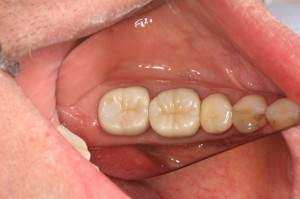
Occlusal view of restored first and second molar.

intentional reimplantation

Side view. The tooth is healthy with no sign of return of the sinus tract and in function.

The three year review PA shows good healing of the apical lucencies. This simple and inexpensive treatment has allowed this patient to keep their tooth in function in the short term. I will keep recalling the patient.

The three year review PA shows good healing of the apical lucency. The lucency associated with the mesial root of the first molar has also resolved. This simple and inexpensive treatment has allowed this patient to keep their tooth in function in the short term. I will keep recalling the patient.

Lets Hear Your Thoughts:

I’d like to hear from others on your technique and experiences with IR. Feel free to send through cases and I’ll add them to this post.

References

Bender IB, Rossman LE (1993). Intentional replantation of endodontically treated teeth. Oral Surgery, Oral Medicine, Oral Pathology 76(5):623-630

Peer M (2004) Intentional replantation – A last resort treatment or a conventional treatment procedure? Nine case reports. Dental Traumatology 20:48-55

The EndoSpot Study Guide on Intentional Reimplantation:

The best known reference for IR is Bender IB, Rossman LE (1993). Intentional replantation of endodontically treated teeth. Oral Surgery, Oral Medicine, Oral Pathology 76(5):623-630. In a retrospective case series they followed 31 cases for  1 day to 22 years and reported an overall success rate of 80%.

Another good overview is provided by Peer, however this is a simple case series. Criteria for success/failure is not listed and no rates of success are given: Peer M (2004) Intentional replantation – A last resort treatment or a conventional treatment procedure? Nine case reports. Dental Traumatology 20:48-55

Primary Indications for IR are listed as:

  1. Teeth have been treated unsuccessfully by conventional means (surgical/non-surgical)
  2. Separated instruments cannot be bypassed or removed
  3. Post/crown restoration requires retreatment
  4. Apical surgery would remove enough bone to cause a periodontal pocket
  5. Apical surgery would result in nerve injury or perforation of the maxillary sinus

Secondary Indications:

  1. To gain access to perforations on the mesial/distal or lingual root surface (esp inraradicularly)
  2. To treat grossly overfilled conventional root fillings and teeth with blunderbuss apices
  3. To enable inspection of a root for fracture
  4. When the patient will not accept apical surgery
  5. When the wrong tooth is extracted or accidental avulsion of a tooth during the removal of crown in a prosthetic procedure;
  6. Cases involving a deciduous tooth to allow it to act as a space maintainer

Contraindications for IR are:

  1. Presence of periodontal disease in which there is marked tooth mobility, furcation involvement or gingival inflammation
  2. Flared or curved roots
  3. Fractured Roots
  4. Patients are taking bisphosphonates

Advantages:

  1. Less invasive than apical surgery
  2. Simpler, and less equipment required c.f. apical surgery
  3. Improved access for inspection and resection/retropreparation
  4. Less soft tissue scarring

Disadvantages:

  1. Potential for root resorption
  2. Reduced access to apical pathology