Aetiology of Apical Periodontitis, Endodontic Surgery

Sometimes, The Simplest Option is the Best Option

When we are dealing with apical pathology, we should always have in our mind that intra-canal infection is by far the most common cause of disease. In a case such as the one shown below, no canal can be seen radiographically, and no canal was found by the dentist that accessed the tooth. But we should remember that a pulp that has undergone pulp canal obliteration will still in most cases have either vital or infected tissue contained within it at a microscopic level.

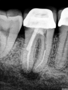
This tooth underwent apicectomy twenty years ago after an unsuccessful attempt to locate a canal. The tooth remained asymptomatic until symptoms of an abscess occurred.

This tooth underwent apicectomy twenty years ago after an unsuccessful attempt to locate a canal. The tooth remained asymptomatic until symptoms of an abscess occurred.

The patient above presented to me for treatment of acute apical abscess. The central incisor had been accessed twenty years earlier, but no canal could be found (despite the large access). At that stage an apicectomy was performed with an amalgam retrofilling placed. The tooth remained asymptomatic for twenty years prior to the abscess forming.

Consider the options we have available:

1. Endodontic treatment, with or without apicectomy;

2. Re-apicectomy;

3. Extraction and bridge/implant.

The previous apicectomy, pulp canal obliteration and large access make this tooth appear to be a difficult to manage case, and this could be mistaken for it having a poor prognosis. But we have to be careful not to over-think the case. With a microscope available, endodontists expect to be able to prepare canals in most teeth such as the one shown above, and therefore take care of the intra-canal infection.

One option would be to take a CBCT to further assess the presence of a canal. In this case however, there has already been an access made, and we are assuming the canal is infected and necrotic, so the decision was made to remove the restoration to see if any canal could be located. The canal was located, prepared and dressed with calcium hydroxide. No exudate was produced, and there was a definite stop at the level of the amalgam retrofilling, The symptoms resolved and four weeks later the canal was filled. The patient was advised that an apicectomy may be required if symptoms recurred. The presence of the amalgam apically, is not by itself sufficient not make apicectomy an obvious option.

Two year review revealed the tooth remained asymptomatic, and healing of the apical pathology had occured. This was despite the amalgam retrofill remaining.

Two year recall. The tooth is asymptomatic and resolution of there is no longer an apical finding present radiographically.

Two year recall. The tooth is asymptomatic and there is no longer an apical finding present radiographically.

The main point to take out of this is that treatment of intracanal infection should be our first priority. Secondly, when we manage to get rid of intracanal infection, then the material used to fill the canal isn’t so important, as long as we maintain a good coronal seal to prevent reinfection. Thirdly, when you see a calcified canal such as this one, it may be a good idea to refer to someone who has experience in these types of cases if this is an option, as the location of the remnant of the canal can be challenging.

Pat Caldwell


Aetiology of Apical Periodontitis, Clinical Tips, Diagnosis of Pulpal Pathology

Viral Pulpitis. This Would Have to Hurt…..


We usually blame bacteria for causing pulpitis but………. This patient complained of severe irreversible pulpitis symptoms for four days, which were only just starting to settle. She couldn’t isolate to either the upper second premolar or molar. Her dentist had extirpated the molar but symptoms hadn’t changed. Pulp testing showed the premolar was also responding negatively.

Take a look at the image below. This presentation is consistent with herpes zoster, commonly referred to as shingles. It is caused by the varicella zoster virus, also responsible for chicken pox. The trigeminal nerve is an unusual location for an outbreak (approximately 1-2% of cases), but when it occurs the virus affects a particular dermatome. If this dermatome also includes teeth, the pulp can also be affected. I can only imagine how painful this would be. Long term follow up is required as multiple teeth may lose vitality.

Notice how the viral outbreak is only affecting the greater palatine nerve distribution. If the nerve innervating the teeth are involved, this can lead to pulpitis and necrosis of the pulp tissues.

Diagnosing this initially may prove difficult, as the lesions on the mucosa don’t show up until a few days after symptoms appear, but some things to look out for include pulpitis symptoms from multiple teeth and a tingling or burning sensation in the distribution of the trigeminal nerve.

Pat Caldwell

Aetiology of Apical Periodontitis, Endodontic Surgery

Choosing your battles – Apicoectomy

I had a patient present to me this week complaining of pain and swelling associated with a lower molar. She had previously had root canal therapy and then apicoectomy performed by her dentist about 8 months ago. She was disappointed that symptoms had recurred as she had seen the dentist in her home country one month ago and he had told her the treatment had been successful, healing was proceeding well and that a crown could be placed. She told me that her dentist had told her prior to the surgery that she had a large lesion and that the actual surgical procedure had been very painful.

apicectomy

Tenderness to percussion or palpation, pain or fistula formation may indicate failure of an apicectomy.

This case is a good one to base a discussion on case selection for apicoectomy. The first thing to note is that the quality of the original RCT is inadequate. One mesial canal is filled well short of the appropriate length. Additionally, the distal canal is poorly filled. There is also evidence that the composite resin restoration may be leaking. It’s reasonable to assume that the root canal was failing due to infection within the canals, and it has been clearly shown that this is the case for the vast majority of root canal treatment failures.

So from a purely endodontic point of view (disregarding alternative options or the influence of prosthetic factors), we have two main options. First, we treat the infection in the canal by undertaking RCT re-treatment. Secondly, we “entomb” the microorganisms in the canal and prevent them and their products reaching the periapical area by undertaking periapical surgery with retrofill.

In this case, we can see that periapical surgery has been undertaken, but no retrofill has been placed. Given the (very safe) assumption that infection remains within the canal, then simply resecting the apical portion of the root will not achieve anything of use. The canal will remain open and the microorganisms will continue to thrive within the canal continue to release products that induce an immune response in the periapical tissues. Unfortunately for this patient, their dentist has shown a lack of understanding of the cause of periapical pathology and needlessly put the patient through a surgical procedure. There may have been some healing of the lesion sue to the surgical procedure, but complete healing could not be expected.

A recent randomized clinical trial compared the outcome of apical surgery where either an MTA retrofilling was placed or the GP filling of the resected root was smoothed and no retrofill placed (Christiansen et al. 2009). The outcome was quite clear that placement of MTA retrofill provides a superior rate of healing compared to just leaving the GP intact. The study didn’t really go into detail as to the quality of the orthograde root fillings in the teeth, but in a situations such as the case discussed above, a retrofill is a necessary part of the procedure. For those of you who don’t have experience with preparing and placing retro fills, it is a skill that requires some practice and specialised equipment. This is especially so once you start to treat molars and teeth with isthmuses.

Without seeing the initial radiographs, I’d have to say that from a purely endodontic point of view, RCT re-treatment should have been considered prior to any surgery. This may have been successful in treating the infection alone, but if not, it provides a better chance of the canals being cleared of infection prior to the surgical procedure, and this then improves the chances of success of the subsequent  surgery. Alternatively, apical surgery with retrofill performed by a skilled clinician might have been successful in this case.

Reference:

Christiansen R, Kirkevang L-L, Hørsted-Bindslev P, Wenzel A. Randomized clinical trial of root-end resection followed by root-end filling with mineral trioxide aggregate or smoothing of the orthograde gutta-percha root filling – 1-year follow-up. Int Endod J, 42, 105–114, 2009.

Aetiology of Apical Periodontitis, Microbiology, Study Guides

The Lazy Man’s Guide to Persistent Apical Periodontitis

Poorly Prepared, Obturated and Missed Canals All Contribute to Persistent Apical Periodontitis

Persistent Apical Periodontitis (AP) refers to AP that is associated with a tooth that has had root canal therapy (RCT).  As with primary AP, bacteria are the most common cause of the inflammatory response (Sudqvist et al. 1998). Previously there has been a large body of evidence that persistent infections are commonly composed of a single species, however recent evidence points to the presence of a mixed biofilm (Siqueira et al. 2009a, Chavez de Paz 2007).”]

There are also non microbial causes of AP, including foreign body reactions, cystic formation, endogenous cholesterol crystals and scar formation. These will be discussed later.

The microbes that cause persistent AP are more commonly located intraradicularly (inside the root). Occasionally, these microbes will also be located extraradicularly. We’ll discuss the far more common intraradicular microbes first.

Intraradicular Microbes

The key study for referencing the presence of microbes within the root in cases of persistent AP is Nair et al. 1990a. When considering the cause of the persistent infection, consider that the microbes were either present prior to RCT being initiated (primary infection ) or they entered during or after treatment (secondary infection) (Siquiera 2008).  When thinking about those microbes that have survived from the primary infection, consider how they might have achieved this. They may have been resistant to the chemicals used in the disinfection process (e. feacalis for example has some mechanisms to survive calcium hydroxide), or they may have been located in a portion of the canal that was not instruments nor cleaned via chemical means.

When considering the secondary infection, these microbes may have gained access to the canal during treatment or after treatment. Think about this also. They may have been carried into the canal on a contaminated instrument or perhaps a leaking rubber dam may have allowed saliva to contaminate the root canal. Alternatively, a poorly placed temporary restoration may have allowed leakage into the root canal system in between visits. If caries have not been completely removed, or a previous restoration which is subject to microleakage is left in place, then this can also be a source of secondary infection. Alternatively, these microbes may have entered a previously clean  root canal system after the completion of RCT. This could be due to a leaking restoration, or through caries or a crack in the tooth. It’s important to understand the microbial nature of AP, and to have this foremost in our minds when undertaking treatment.

Which microbes are present in secondary in persistent AP?

When we look at the composition of the infection in AP, we find a significantly different microflora than that found in primary infections (Figdor et al. 2007, Molander et al. 1998). Generally in persistent AP, there are only 1-5 species.  These are predominantly gram-positive and there is an equal amount of obligate and facultative anaerobes. (Figdor et al. 2007, Sundqvist et al. 1998, Siqueira et al. 2009b). Due to the fact that obligate anaerobes are easier to kill, it may be that facultative anaerobes are more likely to persist within the root canal system after treatment.

E. Faecalis & c.albicans

Enterococcus faecalis is an opportunist pathogen which is implicated in many general surgery post-operative infections.  It has been identified as an opportunistic pathogen in persistent AP in a number of studies (Sundqvist et al. 1998, Sundqvist et al. 2003, Molander et al. 1998). This particular microbe has been studies extensively. It possesses a “proton pump” on its cell membrane which allows it to regulate its internal pH. This means that it is resistant to calcium hydroxide and this may be one of the ways that it survives and becomes implicated in persistent infections. It is also able to survive by itself and without nutrition for long periods of time. It is rarely found in untreated canals. Candida albicans (a fungus) is also found more commonly in persistent infections than in primary infections. (Sundqvist et al. 1998, Nair et al. 1990a, Waltimo et al. 2004)

Extraradicular Infections

Occasionally, we may find a situation where microbes establish themselves outside the root canal system. The microbes may establish themselves on the external root surface in a biofilm, in association with infected dentine chips that have been displaced into the periapical region, or within a periapical cyst (Abbott 2004, Nair 2008). These microbes must be able to withstand the body’s attempts to kill them and it is likely that biofilm formation allows this (Noiri et al. 2002). Similarly in the periapical cyst situation, it is the cyst itself that protects the microbe from the immune response.

In particular, two microbes have been implicated in extraradicular infections. These are Actonomyces spp. And Proprionibacterium proprionicus (Siqueira 2002). These microbes are able to form cohesive colonies within an extracellular matrix. This helps them to avoid phagocytosis, and so continue to survive and invoke the immune response.

Non-microbial causes of AP – Cysts, Foreign Body Reactions and Cholesterol Crystals

In some cases, AP may not be “maintained” by microorganisms. I say “maintained”, because often the AP is initially caused by microbes, and after endodontic treatment, one of the following factors takes over, maintaining the immune response and thus, AP.

Periapical cysts are an interesting topic. There are a range of studies that attempt to measure the incidence of periapical cysts in examined PA lesions. In simple terms, the lesion is biopsied and then examined under a microscope. If an epithelium lined sack is found, then the lesion is designated a cyst. But…… In 1980 Simon published a paper which included serial sectioning of periapical lesions (Simon, 1980). What he found was that some lesions that appeared as cysts on one section, appeared differently on other sections. Thus, it was deemed that the majority of studies (which don’t use serial sectioning) relating to the prevalence of cysts were subject to error. If you just take a random slice, the effect in two dimensions may be that of a cyst, when in reality the full three dimensional structure of the cyst does not exist. Nair repeated this study 16 years later and confirmed Simon’s findings (Nair, 1996).

Nair studied far more lesions than Simon, and found that 15% could be classified as cysts (including both true and pocket varieties). This is probably the best figure to be quoting. Other studies report figures from 5-55%, but they failed to use serial sections. It is also important to realize that a great number a large proportion of abcesses and granulomas will also contain epithelium. In Nair’s study, 52% of the lesions were epithelialised, but only 15% were cysts.  It is likely that the inflammatory process results in the proliferation of this epithelium, and over time, the epithelium develops into a cyst.

Through both of these studies, Simon and Nair found two distinct types of cysts. Simon called them true cysts, those with a complete epithelial lining, and bay cysts, those whereby the lining is attached to the root surface and the contents of the root canal contiguous with the contents of the cyst. Nair referred to these as true cysts and pocket cysts (equivalent to Simon’s Bay cyst).

Nair contends that these two types of cysts are quite different (Nair 2008). He feels that the true cyst is self sustaining, and will remain independent of efforts to remove the microorganisms from the root canal system. The pocket cyst, on the other hand is being sustained by the microbes within the canal system. Removal of the microbes which are maintaining the inflammatory response may allow the pocket cyst to heal. In reality, it will be very difficult to prove or disprove this theory, but one could say that it makes sense.

Foreign body reactions

When exogenous materials are located in the periapical region, they can induce and maintain an inflammatory response which may be asymptomatic, but will be seen as a radiolucency. Materials may be GP, amalgam, sealants, Calcium hydroxide or cellulose fibres such as those contained in paper points (Nair et al. 1990b).

In practice these lesions are rarely seen but have been reported in the literature, so it is important to understand that this mechanism for the maintenance of AP does exist. It also reminds us to be careful when using paper points not to extend them into the periapical areas, as human cells cannot degrade cellulose and leaving fibres behind may result in a foreign body reaction.

Gutta percha may also induce a foreign body reaction, especially in fine particles (Sjögren et al. 1995). Overextended GP may, as a result cause delayed healing of periapical tissues.

Cholesterol Crystals

Cholesterol crystals are also seen in AP, and are probably released by disintegrating erythrocytes, lymphocytes, macrophages, plasma cells and from circulating plasma lipids (Nair 1999). These collections of cholesterol are referred to as cholesterol clefts and induce a reaction similar to a foreign body reaction as the macrophages and giant cells are unable to remove the cholesterol. Again, this may result in a non-healing lesion, despite well completed endodontic treatment.

The Endospot Easy Study Guide to Persistent AP

  1. Persistent AP is most commonly caused by microbes remaining within the root canal system (sundqvist et al. 1998)
  2. It appears that a mixed biofilm may be responsible, contrary to the previous belief that usually only one microbe was responsible.  (Chavez de Paz 2007)
  3. The microbes are either (Siquiera 2008):
    1. Primary – remained within the canal from the initial infection
    2. Secondary – entered during or after treatment
  4. Significantly different flora to primary AP (Molander et al. 1998)
    1. 1-5 species per canal
    2. predominantly gram positive
    3. equal number of obligate and facultative anaerobes
  5. E. faecalis – opportunist pathogen which has been identified more commonly in persistent AP (Sundqvist 1998)
    1. Posesses a “proton pump” which allows it to survive in high pH (i.e. can survive calcium hydroxide)
    2. Can survive in mono-infection
    3. can survive long periods of low/no nutrition
  6. Candida Albicans also found more commonly in persistent infections than in primary (Waltimo et al. 2004)
  7. Exraradicular infections can occur in biofilm on the root tip (Noiri 2002), or in the PA area itself (Siquiera 2002)
    1. Proprionibactrium proprionicus and Acinomyces species are able to from adhesive colonies in an extracellular matrix in the PA tissues
  8. Non-microbial causes of AP are:
    1. Periapical cysts – 15% of lesions (Nair 1996)
      1. Serial sectioning indicates two types – true cysts and pocket cysts
    2. Foreign Body Reactions
    3. Cholesterol Clefts

References:

ABBOTT, P. V. 2004. Classification, diagnosis and clinical manifestations of apical periodontitis. Endodontic Topics, 30-54.

CHAVEZ DE PAZ, L. E. 2007. Redefining the persistent infection in root canals: possible role of biofilm communities. J Endod, 33, 652-62.

FIGDOR, D. & SUNDQVIST, G. 2007. A big role for the very small–Understanding the endodontic microbial flora. Australian Dental Journal, 52, 38.

MOLANDER, A., REIT, C., DAHLEN, G. & KVIST, T. 1998. Microbiological status of root-filled teeth with apical periodontitis. Int Endod J, 31, 1-7.

NAIR, P. N., SJÖGREN, U., KREY, G., KAHNBERG, K. E. & SUNDQVIST, G. 1990a. Intraradicular bacteria and fungi in rootfilled, asymptomatic human teeth with therapy-resistant periapical lesions: a long-term light and electron microscopic follow-up study. J Endod, 16, 580-8.

NAIR, P. N., SJÖGREN, U., KREY, G. & SUNDQVIST, G. 1990b. Therapy-resistant foreign body giant cell granuloma at the periapex of a root-filled human tooth. J Endod, 16, 589-95.

NAIR, P. N. 1996. Types and incidence of human periapical lesions obtained with extracted teeth. Oral Surg Oral Med Oral Pathol Oral Radiol Endod, 81, 93-102.

NAIR, P. N. 1999. Cholesterol as an aetiological agent in endodontic failures–a review. Aust Endod J, 25, 19-26.

NAIR, P. N. 2008. Pathobiology of Apical Periodontitis. Essential Endodontology. 2nd ed. Oxford: Blackwell Munksgaard.

NOIRI, Y., EHARA, A., KAWAHARA, T., TAKEMURA, N. & EBISU, S. 2002. Participation of bacterial biofilms in refractory and chronic periapical periodontitis. J Endod, 28, 679-83.

SIMON, J. H. S. 1980. Incidence of periapical cysts in relation to root canal. J Endod, 6, 845-8.

SIQUEIRA, J. F. 2002. Endodontic infections: concepts, paradigms, and perspectives. Oral Surg Oral Med Oral Pathol Oral Radiol Endod, 94, 281-93.

SIQUEIRA, J. 2008. Microbiology of Apical Periodontitis. Essential Endodontology. Oxford: Blackwell Munksgaard.

SIQUEIRA, J. F., JR. & ROCAS, I. N. 2009a. Community as the unit of pathogenicity: an emerging concept as to the microbial pathogenesis of apical periodontitis. Oral Surg Oral Med Oral Pathol Oral Radiol Endod, 107, 870-8.

SIQUEIRA, J. F. & RÔÇAS, I. N. 2009b. Diversity of endodontic microbiota revisited. Journal of Dental Research, 88, 969-81.

SJÖGREN, U., SUNDQVIST, G. & NAIR, P. N. 1995. Tissue reaction to gutta-percha particles of various sizes when implanted subcutaneously in guinea pigs. European Journal of Oral Sciences, 103, 313-21.

SUNDQVIST, G. & FIGDOR, D. 2003. Life as an endodontic pathogen. Endodontic Topics, 6, 3-28.

SUNDQVIST, G., FIGDOR, D., PERSSON, S. & SJÖGREN, U. 1998. Microbiologic analysis of teeth with failed endodontic treatment and the outcome of conservative re-treatment. Oral Surg Oral Med Oral Pathol Oral Radiol Endod, 85, 86-93.

WALTIMO, T. M. T., HAAPASALO, M., ZEHNDER, M. & MEYER, J. 2004. Clinical aspects related to endodontic yeast infections. Endodontic Topics, 66-78.

Aetiology of Apical Periodontitis, Microbiology, Study Guides

The Lazy Man’s Guide to the Microbial Causes of Primary Apical Periodontitis

R1This is a dry topic, so we’ll try to cover it as simply as possible. If you just want the outline, check out the study guide at the end of the blog post.

So Microorganisms cause Apical Periodontitis. How do they get into the tooth?

Primary apical periodontitis (AP) occurs when microorganisms (let’s call them bugs) enter the pulp chamber and colonise the pulp tissue. The bugs enter the pulp chamber through a variety of routes such as carious lesions, cracks, traumatic exposure of the pulp and due to dental procedures such as cavity preparation (Nair 1997). As mentioned in the previous post on the aetiology of AP, there is plenty of evidence of bugs being found in teeth that have apparently intact crowns (Bergenholtz 1974). These bugs gain access through accessory canals, exposed dentinal tubules, or microcracks adjacent to the gingival crevice or deep periodontal pockets (Nair 1997). The important message here is that we don’t require an obvious,visible path of entry to the pulp chamber. Bugs can enter through tiny, microscopic spaces such as dentinal tubules.

Continue reading

Aetiology of Apical Periodontitis, Study Guides

The Lazy Man’s Guide to the Aetiology of Apical Periodontitis

Lateral Incisor with Apical Periodontitis

What's the Root of This Problem?

Generally, apical periodontitis (AP) is a result of microorganisms entering the root canal system. Of course there are other causes of inflammation in the apical area, such as trauma, traumatic occlusion and foreign body reactions, but if we’re talking about common AP, then “bugs” are the cause (Abbott 2004, Harn et al. 2001).

The classic study on this topic was Kakehashi et al (1965), and this should be the starting point for any essay or exam answer. They took gnotobiotic rats (bacteria free rats) and conventional rats (with bacteria) and exposed the pulps in some teeth. In this study, the germ free rats had food, and in one case a hair forced into their long term exposed pulps, but necrosis did not occur! There was even dentine bridge formation in some cases. Conversely, all the conventional rats developed pulpal necrosis and apical periodontitis. It seems that without bacteria, rat pulps are very hardy indeed.

Sundqvist (1976) was the first to anaerobically culture and taxonomically analyse the constituent bacteria in teeth with necrotic pulps. He concluded that: AP is associated with the presence of bacteria in the root canal and that infected teeth harboured a mixture of bacteria that were predominantly obligate anaerobes (Sundqvist 1976). The significance of obligate anaerobes in endodontic infections are confirmed by a number of independent studies (Bergenholtz 1974, Wittgow et al. 1975, Kantz et al. 1974).

A study by Möller et al. (1981) compared the apical response of inducing pulp necrosis both aseptically and after contamination with indigenous oral flora in monkeys. Basically, they aseptically opened monkey pulps and used a Hedstrom file to mince the pulp. They then closed the pulp chamber so that the necrotic, uninfected tissue was inside, or they infected the tissue with plaque form the monkey’s teeth. In the teeth which contained sterile necrotic tissue, no AP formed. Those with bacteria were associated with AP. At this stage there was still a commonly held belief that necrotic tissue and stagnant tissue contributed to AP formation, but this study helps dispel this theory. In addition, this study showed that there was a variation in the ability of the various bacteria to survive and establish themselves within the root canal environment, and we start to see the concept of bacteria within root canals living in communities, rather than just as one individual strain.

There has been a significant shift away from the concept of one ‘causative pathogen’ and significant evidence exists that specificity seems to be related to the community level as certain species compositions are specifically associated with some forms of apical periodontitis (Siqueira et al. 2009a, Sakamoto et al. 2006). There is a very high level of inter-individual variation of bacterial community profiles, such that 44% of recovered taxa were isolated or detected in only one study (Siqueira et al. 2009b). Microbial ecology differs significantly between different disease forms such as chronic apical periodontitis versus acute apical abscess, suggesting the existence of a pattern associated with each one (Sakamoto et al. 2006, Siqueira et al. 2004). In addition, different magnitudes
of disease, based on intensity of signs and symptoms, may be related to the species composition of the community (Siqueira et al. 2009a).

Siqueira (2002) differentiates the types of endodontic infections into primary, secondary or persistent root canal infections.  Primary infections are those that occur within a root canal system prior to any treatment being applied. A Secondary infection occurs when a root canal (RC) had been treated, and new bacteria enter the RC system.  This can happen when a filling is lost, or micorleakage occurs and the root filling (RF) is exposed to bacteria which then penetrate along the RF. A persistent infection is one that remains in the canal despite treatment. That is the primary infection remains despite RCT.

Primary Apical Periodontitis

This molar exhibits apical periodontitis. It has not been endodontically treated. This is an example of primary apical periodontitis.

Persistant Apical Periodontitis

The first molar exhibits AP associated with both roots. The disease could be persistent (remaining after treatment), or secondary due to coronal microleakage.

An important concept to understand is the formation of biofilms. This relates to the organisation of bacteria into a protected, sessile biofilm comprised of cells embedded in a hydrated exopolysaccharide-complex in microcolonies (Nair 2006). Bacteria in root canals do form biofilms and these are much more difficult for both our immune system and current RCT methods to deal with.

Do you have a reference that should be included in this list, or a comment on what is written? Please share it with our readers.

References:

ABBOTT, P. V. 2004. Classification, diagnosis and clinical manifestations of apical periodontitis. Endodontic Topics, 30-54.

HARN, W. M., CHEN, M. C., CHEN, Y. H., LIU, J. W. & CHUNG, C. H. 2001. Effect of occlusal trauma on healing of
periapical pathoses: report of two cases. Int Endod J, 34, 554-61.

KAKEHASHI, S., STANLEY, H. R. & FITZGERALD, R. J. 1965. The Effects of Surgical Exposures of Dental Pulps in Germ-
Free and Conventional Laboratory Rats. Oral Surg Oral Med Oral Pathol, 20, 340-9.

MÖLLER, A. J., FABRICIUS, L., DAHLÉN, G., OHMAN, A. E. & HEYDEN, G. 1981. Influence on periapical tissues of
indigenous oral bacteria and necrotic pulp tissue in monkeys. Scand J Dent Res, 89, 475-84.

NAIR, P. N. R. 2006. On the causes of persistent apical periodontitis: a review. Int Endod J, 39, 249-81.

SAKAMOTO, M., ROCAS, I. N., SIQUEIRA, J. F., JR. & BENNO, Y. 2006. Molecular analysis of bacteria in asymptomatic and
symptomatic endodontic infections. Oral Microbiol Immunol, 21, 112-22.

SIQUEIRA, J. F. 2002. Endodontic infections: concepts, paradigms, and perspectives. Oral Surg Oral Med Oral Pathol Oral
Radiol Endod, 94, 281-93.

SIQUEIRA, J. F., JR. & ROCAS, I. N. 2009a. Community as the unit of pathogenicity: an emerging concept as to the
microbial pathogenesis of apical periodontitis. Oral Surg Oral Med Oral Pathol Oral Radiol Endod, 107, 870-8.

SIQUEIRA, J. F. & RÔÇAS, I. N. 2009b. Diversity of endodontic microbiota revisited. Journal of Dental Research, 88, 969-81.

SUNDQVIST G. 1976. Bacteriological studies of necrotic dental pulps. Odontological Dissertations No. 7. Department of Oral Microbiology, Umea° University, Sweden.

The Endospot Easy Study Guide on Aetiology of Apical Periodontitis

  1. Most AP is due to micororganisms within the RC. Other occasional causes include trauma, occlusal trauma, foreign body reaction. (Abbott 2004, Harn et al. 2001)
  2. Exposing pulps in rat teeth lead to AP in conventional rats (bacteria present), but no AP in gnotibiotic rats (no bugs present) (Kakehashi, 1965)
  3. Bacteria in infected necrotic pulps predominately obligate anaerobes. Anaerobic techniques required to culture (Sundqvist thesis, 1976)
  4. Monkey teeth with necrotic, uninfected pulps do not develop AP. If infected, AP develops. Different microorganisms display differing ability to survive in the root canal system. (Mollar, 1981)
  5. It is unlikely one microorganism causes disease in RCs. More likely that a community of mircobes exists. This community varies between people, and between various forms of disease. (Siqueira et al. 2009a, Sakamoto et al. 2006)
  6. Biofilms form in RCs. Biofilms protect bacteria from being destroyed. (Nair 2006)
  7. Primary infections (new in previously unifected RCs), secondary infections (new in previously treated RCs), persistent (remaining after treatment). Siqueira (2002)