This patient presented with occasional symptoms. There was a large recurrent carious lesion present and radiographically an apical finding on the mesial root. Once caries was removed, the pulp was exposed. It is interesting that the pulp was vital and inflamed. Often, having an apical radiolucency and a vital pulp is confusing, but basic biology explains this phonomenon.
Once the pulp of the tooth starts to become inflamed, for example due to caries, changes start to occur in the apical tissues. This includes the processes that leads to demineralisation of bone. As this process develops, the demineralised bone will become apparent as an apical lucency. The apical bone doesn’t want to wait for bacteria to reach the apical foramen before it starts to remove bone and form inflammatory tissue. It wants to do this well before the bacteria reach bone. Bone has a relatively poor blood supply and doesn’t handle infection well.
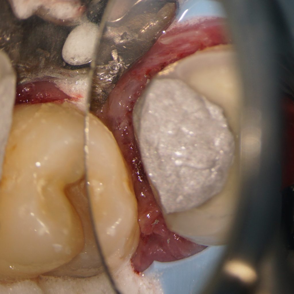
In this case, the endodontic treatment was completed over a single visit as the patient travelled a number of hours for treatment. A gingivectomy was required distally and an amalgam core placed. The contact was left open so the patient can scrub the interproximal area with interproximal brushes prior to crown preparatio.n. A full gold crown has been recommended.