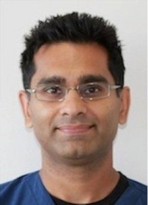
Oliver Pope is an Endodontist practicing and teaching in Melbourne, Australia. He has been involved in researching what “normal” looks like on cone beam CT, and has fantastic insights on the relevance of CBCT findings to everyday practice. In part 1 of this podcast we discuss the interpretation of apical findings on CBCT, and the limitations of the technology, as well as comparing use of PAs with CBCT.

Podcast: Play in new window | Download
Show Notes and Links
Find Oliver’s practice on Collins St in Melbourne
Carestream 9000 CBCT, and the newer 8100 Model
Morita Accuitomo 80 CBCT
The Transcription:
00:08.7
PC: Welcome to the Endospot. On the Endospot we interview world-class endodontists to find out what gives them the ability to achieve things the rest of us are unable to. They will provide knowledge, skills, and techniques that you can use to improve your endodontics everyday. In this episode, I speak to Oliver Pope and pick his brains about cone beam CT. I interviewed Oliver in Brisbane, Australia.
00:30.5
OP: I guess something that I am familiar with is cone beam computer tomography by virtue of the fact that through my post-grad training, my research project was on and my thesis was on CBCT.
00:43.9
PC: So tell us quickly what your project was about.
00:47.1
OP: When I started post-grad, there was a lot of research coming out on CBCT. A lot of the research was showing that CBCT, by virtue of the fact that it is three dimensional, shows a lot more things, obviously, than a two dimensional periapical radiograph. The main thing that we are showing is that there are lesions visible, or bigger, on a CBCT than they are on traditional PAs.
01:09.8
PC: So, for the same pathology, it is more obvious on cone beam than it was on a PA.
01:13.9
OP: Correct. Correct. Because, obviously with 2D image you get a lot of superimposition of anatomical structures. You do not have so much, it is less of an issue with CBCT. Because of all this new information about CBCT, there was a bit of a call that CBCT should be replacing periapicals as our pre-diagnosis and post treatment. Basically replacing PAs. There are arguments for and against that, but one of the fundamental bits of information that we were missing was that what does a healthy tooth look like on a CBCT compared to a PA.
When we started using PAs, there was a lot of research done in the 60s as to what the correlation is between inflammation and radiography and apical periodontitis is inflammation around the apex. The reason we use radiographs is because there is a loose correlation between apical inflammation and what it looks like on an x-ray, that we didn’t have that information on a CBCT at all. It was one small step to see what the apex of a healthy tooth looks like on a CBCT.
Basically, I asked all the local endodontists to submit cases. I spent a lot of time going through their records to find a number of data that had a CBCT of a tooth, a small field of view CBCT, a traditional radiograph, and then a healthy pulp test response. Then we correlated all those, to look at them.
02:45.9
PC: How many cone beam CT would you have looked at?
02:48.6
OP: I probably looked at around 300 teeth. Then, out of that there were 200 that were included because of those three criteria.
02:57.7
PC: Looking at 200 cone beam CTs of healthy teeth, that is a lot of cone beam CTs to look at. Your average dentist or even a lot of endodontists would not have looked at that many cone beam CTs. When you did that project, what are the main things that you found of interest?
03:13.5
OP: Basically, a healthy periodontal ligament looks different on a CBCT than it does on a PA radiograph.
03:21.3
PC: Always?
03:21.9
OP: A vast majority of the time. They always look bigger, the PDL. There is a bigger space of a PDL. When you look at a healthy tooth on a PA radiograph, we know quite confidently there is not going to be a widening of the periodontal ligament, there is not going to be a change in the periodontal ligament space. It conforms to the shape of the root. Whereas CBCT you can’t apply that. In my study I found that only 25% of the teeth looked similar to that no widening. Roughly 50% of the teeth had a widening roughly of up to a millimeter; 25% had widening of one to two millimeters. These are healthy teeth. The inference from that is that if you look at a root-filled tooth on a CBCT that might have a widening of two millimeters, for example, that still could be normal. It is just a difference in how you look at them.
04:13.8
PC: And the risk is, if you are not familiar with looking at cone beam CT, you could look at a healthy tooth and make the assumption that it has some sort of pathology associated with it.
04:23.6
OP: I think something that happens out there in clinical practices is that, of course, CBCTs give these beautiful 3D images, they are much more accurate and better than PAs in certain aspects. But also, they can’t replace clinical pulp testing examination. We have remember that all the time. Those are the fundamental basics, and 90% of our diagnosis comes from the clinical findings.
04:48.2
PC: What do you think are the main challenges we have in reading cone beam CTs or using cone beam CTs?
04:55.3
OP: I think they are two different imaging modalities. We as dentists, ever since second year dental school, we read a lot of bite-wings, PAs, and OPGs. So we are very familiar with that. Even though you might not know it at the undergraduate level, there is a lot of research behind how you read those. You look at those traditional radiographs and then you subsequently treat the teeth. So, you are familiar with the way the appearance is on a radiograph and what it looks like in a clinical scenario.
But that doesn’t directly apply to CBCTs, because most people are not looking at a lot of them, so they knew they were completely different. The way that the image is generated is different, so periapical radiograph is a single beam that passes through all the structures and has various attenuation or absorption, then it ends up as an image on a radiograph. Whereas a CBCT is different because it is a three dimensional image. You get blocking at various points because of radio-dense objects, and then a computer has to actually generate that image. There is a whole number of artifacts that exist in CBCT that don’t exist in periapical, because it gets a lot of different angles from that one blocking. It shifts and takes another image where it is blocked. There is a point whereby it is blocked, so it has to use algorithms to generate that image.
My clinicians do not look at a lot of cone beams and then do not treat from those cone beams. There is that level of connection between the radiography and the clinical scenario. Interproximal caries on a bite-wing…you look at that for awhile on a certain type of x-ray, and then you get very confident on what is caries and what is not caries. Any change by going to digital, you need to rejig that. The same thing is with CBCTs.
06:39.8
PC: When do you think you gain the most benefit from using cone beam CT in your practice?
06:45.9
OP: I think there are two main aspects. Diagnosis of apical pathology on certain roots, and also identification of additional anatomy that is not present or not obvious on a periapical radiograph.
07:00.1
PC: What would prompt you to take the cone beam CT?
07:02.0
OP: I think it is a case-by-case basis. Most of the time, taking a CBCT, is after I have been in the tooth. For example, an upper molar, I am going to treat three canals quite simply, and I am very highly suspect that there is an MB2 canal. Clinically I cannot see it. I have troughed conservatively, I cannot find an incident and I cannot find entrance to the canal, but we know that a massive percentage, 80-90% of the MB2 canals. I am not comfortable operating on a tooth unless I found or I know definitively that there is an MB2 canal present.
07:40.0
PC: Do you ever find on the scan you can see the canal, but then you cannot treat it?
07:45.1
OP: Occasionally. It happens occasionally, when it splits off and is very deep down in the mesial buccal, when there is a big loss of dentine. If you trough five millimeters into a mesial buccal root…I think there is a role for CBCT with general practitioners with endodontics in managing additional canals with this. And when you are looking at a scan for an MB2 canal, a few points I want to make is, it is going to be a small field of view, high resolution scan. There is not really a lot of point in looking at an i-CAT full head to look for an MB2 canal. Sometimes you get lucky and see it.
If you are looking for an MB2 canal, do not refer to a full-head i-CAT. You would be unlikely to see it. When you are looking for the canal on the scan, you are not necessarily going to see a radiolucency, you are not going to see a black spot on the scan because sometimes it is too small to be picked up even by a CBCT. But what you will see is an asymmetry in the presence of the mesial buccal, MB1 canal, so you will see it off to the side of the root, in which you are very confident that there is something on the MB2 side, whether it is an isthmus or a fin or a or canal, and it does warrant further investigation.
08:53.3
PC: When we say we see a patent canal on a cone beam CT, what are we really seeing there? Does that mean there’s a patent canal there?
09:01.8
OP: Not necessarily, because you have to remember, what you are looking at is attenuation of an x-ray beam. So, absorption into that material will show as opacity, but in case of a patent canal, if you see a radiolucency in the canal, it might be filled with resin or a Russian red root-filling paste. Just because you see a patent canal, does not mean that it is actually patent.
The aspects of a CBCT machine, one of the primary things is the field of view. A large field of view obviously scans a large area and irradiates the patient more. A small field of view is focused on a smaller area, and you tend to get a high resolution, and it is also generally less radiation to the patient. Because of that, there is less radiation, because you are only scanning areas that you actually need is certainly more appropriate.
09:55.9
PC: What is the most important aspect about it?
09:58.7
OP: The high resolution, small field of view.
10:00.7
PC: What machines have you used?
10:02.9
OP: There is a handful of machines out there. The ones that I am familiar with most are the Kodak 9000 and the Morita Accuitomo 80. They both give very good images. In my practice in Melbourne, there are two radiology practices that have an Accuitomo 80, one in the CBD Clayray Radiology, another one out at Camberwell called DMDI which is a bit more our in the suburbs. They both give excellent images. But you have to remember it is a small field of view.
10:35.4
PC: They are reported on by an oral radiologist?
10:38.3
OP: They are reported on by an oral radiologist.
10:42.0
PC: Switching tracks a little bit, tell me what do you think that the dentists who are keen to improve their endodontics, what do you think is the most important thing that they can do?
10:47.6
OP: In a general sense; the best thing they can do is to take a bite-wing, in addition to a periapical radiograph, because one of the hottest things I find when treating a tooth is the size of the pulp chamber. If you are treating a tooth that has a really calcified pulp chamber, that highlights to me that it is going to be a very difficult case. Almost irrespective of what the canal is, is finding the pulp chamber. Is it conservative access is one of the hardest things. Also, the bite-wing gives you a map in terms of depth your burr should be at in relation to the crown or the angulation of the tooth. Those are the cases where I see damage is done, and unfortunately we cannot fix. That is probably the greatest thing.
Secondly, once the canals are found, not placing a rotary instrument in there until you have got a really good glide path. Put a small file in first, size six, then you can work it up, not until it is loose with a size 15, should you put something spinning in the canal.
11:53.3
PC: I completely agree. I think the thing that most commonly makes endo difficult is when there is other calcified pulp, all the calcification within the pulp chamber, pulp stains, etc. It is confusing. Even after you have looked at thousands of teeth, it can be confusing when it is calcified, trying to work out where the canals are. Conversely, when there is a large pulp chamber, it generally is easy.
What, then, makes a good referrer?
12:20.5
OP: I think the best referrer is some whose number one concern is high quality treatment to their patients. But also values the network in which they work. That comes from DAs to hygienists to therapists themselves up through the specialists. A similar thing is that they know their limits at which they can provide the highest quality care, but those limits are constantly being expanded in which they are still providing the highest quality of care. The first year graduates are very different from a general dentist at five years and ten years. With time, the scope of your practice is going to improve, but I think the goal of the highest quality of care is most important.
In light of what makes a good referrer, what makes a good specialist is also the same sort of answer; someone who values that network and is aiming to improve the limits of every general practitioner. The role of the specialist is that we lead certain fields so that everyone else comes with us. Educating and being involved in improving the quality of care for general dentists is important. I want my referrers to be the best general dentists in the town. That is the perfect network, really.
13:53.3
PC: How do you go about building that network and an endodontist?
13:57.2
OP: Relationships. The relationship is a two-way street. It comes down to personal, normal, human relationships. I need to feel comfortable to telephone them. Sometimes I have a patient in the chair, and I am unsure about a certain issue or restorability or sometimes I am uncomfortable with something, it is great to be able to pick up the phone and call them to ask them, and get the information straight away and vice versa, so that the dentist feels comfortable identifying a case, or if he is uncertain about whether their case is manageable, asking us. Because we all want the same thing.
14:30.1
PC: And what makes a bad referrer?
14:31.6
OP: What makes a bad referrer is a dangerous question! Basically, someone who does irreversible damage and then continues to do so. Everyone makes mistakes. However, if people continually do that and not acknowledging that they are making mistakes, and they are not open to that communication to improve what they are doing, and improving their clinical skills, that is someone that we do not want to work with.
14:55.4
If a referrer had a problem with treatment you don’t know that they were not happy, how would you invite them to go about dealing with that?
15:02.8
OP: In dentistry, if someone is unhappy with someone, how would you deal with it? You want a phone call, or a letter, or we want to know because it is a two-way street. If something happens that is untoward and we don’t hear about it, we want to know. For instance, someone says that they are getting angry because root canal treatments do not work, because the teeth always break. I hope that if that ever happens that they are telling their specialist that this is happening because we need to know.
15:30.6
PC: We had a lecture recently that covered this topic, and I said that they did a ten-year review of patients. Of the teeth that had been extracted in that patient pool, they did not know of any of them that they had not been informed or they had not reviewed the patient and found out that the tooth had been extracted. That is pretty important, because how to then be looking at the sorts of things that might be associated with failure. If you are not seeing the failures, if a tooth failed, or it was difficult to restore, or it did not feel quite right, or we made a bad call, in any way, it would be grateful to get that sort of feedback.
Going back to post-grad training, what prompted you to become an endodontist?
16:12.5
OP: After uni, I joined the Navy, which was a great thing to do. If anyone thinks they might be remotely interested in a military lifestyle of moving around, getting the experiences that you will never have in another time, is a fantastic thing, and I would do it again in a heartbeat.
16:29.3
PC: What was your favorite experience in the Navy?
16:31.9
OP: I got to fly in and to fly a Hawk Fighter jet in southern England. I got to know one of the pilots and he asked me if I wanted to go with them while they were doing a training operation. I was flying in the navigator’s seat. On the way back he gave me the controls to fly the jet. That was an incredible experience.
During the navy we have an official mentorship program that happens with the senior dentist or specialist. I had an endodontist that was mentoring me. In the practice I was at, we had a microscope. When the microscope was not being used by a visiting an endodontist, I got to use it. With time, I realized that I wanted to focus more on that, and then apply it to post-grad study. I was lucky enough to get in.
17:22.4
PC: What do you have to do to get into endodontics? You trained in Melbourne, right?
17:25.4
OP: Yes, I trained in Melbourne. You need to work at this if it is really what you want to do. Doing endodontics, you do root canal treatment all day. Occasionally it is dispersed by apical surgery or post management of trauma, but mostly it is root canal treatment. As a general dentist, one of the great things is you can do a wide variety of things, and you can focus on things you like, and not on the things you do not like. You can adjust your practice accordingly. There are benefits of being a general dentist. If you think you are interested in endodontics, you have got to make really sure that is what you want to do, because it is sometimes hard to answer to a non-dental person as to what you do. You tell them you are an endodontist and do root canal treatments. And they ask what else you do…and then the conversation is over!
The best way to make sure you want to do it is to spend time with endodontists. See what they do day-in-and-day-out.
18:20.6
PC: Do you think you have to be a little bit weird to want to be an endodontist?
18:23.9
OP: I would think so.
18:29.5
PC: How did you get into the program?
18:30.9
OP: I did the fellowship as far as CDS primaries, training coach, and then applied to endo in Melbourne, not expecting to get in the first year. But maybe by a stroke of luck, I got in after a series of interviews.
18:47.9
PC: Any tips for people who are considering going through that process?
18:51.4
OP: You have got to show that you are focused on doing endodontics. You also have to show that you have a wide general practice background. Even though you want to focus on endo, you have got to do other things. Because, in the post-grad training program, you get taught how to do endo, but among the other difficult things through the course is the other general practice treatment planning issues. You have to be strong on that. Knowledge when going into a specialist training interview, you have to show that you came, but also show that you do not necessarily know it all. From a program point of view, they want someone they can train and has an opened mind. What you learned may not be the way that the school teaches.
19:31.6
PC: What was a difficult aspect of doing your post-grad training? Or did you find it difficult? Did you enjoy it?
19:37.1
OP: I enjoyed it. But the most difficult thing was the time. The time you have to apply. Remember, you spend most of your day in clinic, and then afterward you might have emergency service or after hours paying service you have to do a certain number of rotations. Then after that you have to write seminars, you have to write lectures, do literature reviews. It is relentless! At the time, you get told by other endodontists and specialists that this is a great time in your life and to really enjoy it, but by that time you think you are in the burning pit of hell. However, at the same time, it is a very enjoyable process.
20:16.6
PC: I thought it was fun developing the skills. It was fun being in the uni environment.
I think that is all for today, we can follow up with a part two at some time if you will come back.
20:29.8
OP: Or maybe you need to come down to Melbourne.
20:31.9
PC: That would be even better. Thank you very much for giving us your time and giving us your wisdom as well. We will look forward to seeing you next time.
20:38.8
OP: Thanks then.


