General Updates, Podcast

Podcast Episode 4 – Viraj Vora on Making Endodontic Practice Successful

Dr. Viraj-Vora-Headshot

Dr Viraj Vora is an endodonist in Toronto, Canada. He is known as a critical thinker, and a talented clinician. In the episode he shares with me his thoughts on what makes an endodontic practice successful and also some of the thought processes he goes through when assessing cases. This episode is essential listening for endodontic residents and endodontists starting out in practice.

iTunes

Show Notes and Links

Find Viraj’s practice in Vaughan, Toronto, Canada

Follow Viraj on Facebook

Vortex Blue endodontic files

Cracks in Teeth, General Updates

12 Month Recall on Lower Molar with J-Shaped Lucency

It’s important to confirm the presence or absence of a crack in these teeth before consigning them to extraction. There was no obvious crack or periodontal probing. The lucency was endodontic in origin only.

Pre-op. J-shaped lucency and draining sinus present.

Pre-op. J-shaped lucency and draining sinus present.

Post-op. 4 weeks with calcium hydroxide and the sinus tract closed.

Post-op. 4 weeks with calcium hydroxide and the sinus tract closed.

12 month recall.

12 month recall.

In this case the radiographic image was typical of a root fracture. However without confirming a fracture visually, the radiographic image alone is insufficient to decide a prognosis for the tooth.

Clinical Tips, Endodontic Preparation Techniques, General Updates

The Lower Second Molar. Tricky in So Many Ways.

This case was referred as the treating dentist was astute enough to recognise that while some lower second molars only have two canals, the majority will have at least three. Only one orifice was visible mesially. The mesial roots of lower second (and third) molars are often challenging to treat for a number of reasons. Access can be difficult, there is often a double curve in the mesio-distal and bucco-lingual plane, and often we see a single orifice with two canals. This was one of those cases where only a single orifice was visible mesially.

Before attempting to tackle this case we took a CBVT. The CBVT presented a finding indicative of a second canal in the mesial root. This is good information to have, as if we suspect there are two canals, we know where to remove dentine in an attempt to locate the canal. If we suspect only one canal after viewing the CBVT, we don’t need to be needlessly removing further dentine in the all-important pericervical area.


The clinical picture is of one mesial orifice, but the CBVT indicates two canals.

The clinical picture is of one mesial orifice, but the CBVT indicates two canals.

The CBVT also tells us where to look – buccal or lingual of the orifice. In my experience, the classic situation for these canals is for the main canal to be the mesiobuccal, with the mesiolingual canal coming off the main canal a few millimetres below the orifice. The orifice to the second canal can be quite small, and as it comes off the main canal at a sharp, almost 90 degree angle, we have to be careful to open the canal coronally with some hand files before attempting to place a rotary file in it.

After removing about 2mm of dentin lingually to the existing orifice, the second canal was located.

After removing about 2mm of dentin lingually to the existing orifice, the second canal was located.

The final root filling with composite closing the access.

The final root filling with composite closing the access.

So there you have it. One more little gem in your bag of tricks for treating difficult anatomy. Have you had experience in cases such as this? Share your comments below.

Pat

General Updates

Cases of Note: Silver Point Removal

The anterior abutment of this bridge was symptomatic. The patient was keen to retain the bridge if possible. A decision was made to attempt re-treatment thorough the bridge.

Three silver points were visible after removal of the core material.

Three silver points were visible after removal of the core material.

One of the keys to removing silver points is to carefully dissect the core material around the point so that the end of the point can be grasped with Steiglitz forceps.

The silver points were grasped with Steiglitz forceps and removed.

The silver points were grasped with Steiglitz forceps and removed.

The tooth was dressed with calcium hydroxide for two weeks, symptoms resolved and obturation completed with GP and AH Plus. Two fibre posts were placed with a luxacore core. The porcelain was etched and silanated, and opaque porcelain was used to help mask the darkness of the metal base and the access restored with composite. Some of the porcelain had fractured away during access and this was restored with composite as well.

Obturation with two fibre posts and a luxacore core.

Obturation with two fibre posts and a luxacore core.


Post-obturation radiographs.

Post-obturation radiographs.

Would you have managed this differently? If so, make a comment below.

General Updates

Longer Lasting Endo. Is Smaller Stronger?

Recently there has been a push towards patient-centred outcomes in dentistry. This means we should treat towards outcomes that are important to the patient. Most patients want teeth that are aesthetic and free of disease and pain, and they want the teeth to be there forever.

In recent years, there has also been a push towards conservation of dentine during endodontic procedures in order to maintain as much strength in the tooth as possible. A recent study by Krishan et al showed that the concept of “straight line access”  may lead to significant decreases in fracture resistance in premolar and molar teeth. Although this is a bench-top study, and a long way from a definitive link to outcome, to me it makes sense that if we are going to give the patient the longest lasting solution, then giving them the strongest tooth possible should be one of our primary goals.

In this case, a 22 year old patient was referred for endodontic treatment of the lower left second molar as an odontogenic cyst was to be removed and this would have resulted in loss of vitality of the tooth. There had also been some resorption of the mesial roots. Careful planning of the access allowed all canals to be treated without unnecessary removal of of “pericervical” dentine, the tooth structure that Clark and Khademi feel is the most important to be conserved. The pulp chamber was filled with amalgam and the access closed with composite resin.

I would warn that this approach is more clinically challenging to do well, and that when attempting to be conservative, that the procedure should be titrated. That is, if you need to prepare a canal, and your access isn’t allowing you to do this, then obviously, you need to remove further tooth structure in order to complete the endodontics.

Endodontic access allows good vision and file access whilst conserving tooth structure.

Endodontic access allows good vision and file access whilst conserving tooth structure.

Pulp chamber filled with amalgam and the access closed with composite resin.

Pulp chamber filled with amalgam and the access closed with composite resin.

References:

Krishan R, Paque F, Ossareh A, Kishen A, Dao T, Friedman S. Impacts of Conservative Endodontic Cavity on Root Canal Instrumentation Efficacy and Resistance to Fracture Assessed in Incisors, Premolars, and Molars. JOE In Press.

Clark D, Khademi J. Modern molar endodontic access and directed dentin conservation. Dent Clin North Am 2010;54:249–73.


Diagnosis of Pulpal Pathology, General Updates

Overdiagnosis in Endo? Surely not!

I’ve noticed that the issue of overdiagnosis is really getting a lot of attention in the medical world. Overdiagnosis occurs when a person is diagnosed with a disease, but that disease, if left untreated would never have caused symptoms or death. This results in treatment being provided to people when they could have been left alone and never would have suffered. It is especially problematic when testing is involved that returns abnormalities that can be interpreted to indicate disease, but the test is inaccurate. It is also especially problematic when the treatment has significant consequences or side effects.

There was a conference held on the topic last year in the UK and I see this year’s conference is sold out. httpv://www.preventingoverdiagnosis.net/

I must admit that after reading a bit on the topic, there are a lot of aspects of overdiagnosis that apply to dentistry, as we are so focused on prevention of disease, and we do a lot of tests (pulp testing, bitewing and PA radiographs, probing of fissures) that can return both false positives and false negatives. Also, our treatments are invasive, costly and often irreversible.

This youtube video gives an easy to understand overview of testing: https://www.youtube.com/watch?v=U4_3fditnWg.

What are people’s thoughts on this? Do you think we are guilty?

General Updates, Study Guides

Are Most Published Research Findings False?

Slimey DelightAlong with some other enlightening speakers, I just had the pleasure of listening to a full day of lecture material by Dr John Ioannidis. Dr Ioannidis is a leader in the field of Epidemiology and Research Statistics. In 2005 he authored an article entitled, “Why Most Published Research Findings are False”.

I’ve read his articles before, but having the man himself run through each of the areas covered in the article was a real eye opener. I don’t want to be critical of all the dedicated and hard-working researchers out there. We owe them a debt of gratitude. But, it would seem that the publishing of research findings is a an area where accidental (or non-accidental) misleading statement, conclusions and assumptions are often made.

The biggest issue we have is the pursuit of findings that are often based on research with a formal statistical significance, commonly with a p value less than 0.05. Dr Ioannidis shows that these findings can often be incorrect. He should know. He’s been involved in large scale studies which made findings that were later refuted and shown to be (more than likely) incorrect.

As practitioners, we are called upon ever day to make decisions based on the published research, so it is important that we have a good understanding of what we are reading. I recommend reading the article by Dr Ioannidis and taking note of the corollaries he mentions. It is this sort of introspection which makes us better practitioners.

Please, read his article. In fact, don’t read it, study it, and get interested in what all those statistics actually mean.

John Baez does a nice job of simplifying the corollaries, and there is some good discussion to follow it on his blog.

The article is free here: http://www.plosmedicine.org/article/info:doi/10.1371/journal.pmed.0020124

Conservation of tooth structure
Clinical Tips, Endodontic Preparation Techniques, General Updates

Endodontic Access for Conservationists. A How To Guide.


There are a huge number of factors that can affect the outcome of endodontic therapy. Attempting to isolate these individual factors and determine the relative importance of each is something that has proven difficult or impossible in endodontic scientific literature.

Ultimately, our primary aim is to allow our patients to keep their teeth for the rest of their lives. The evidence we have available points to the majority of endodontically treated teeth surviving for long periods of time. (1) Those that are extracted most commonly fail due to non-endodontic reasons. The most common cause of extraction of root filled teeth is crown fracture and periodontal disease.(2)

In terms of crown (and root) fracture, I believe that the conservation of dentine in the crown and in the region a couple of mm just above and below the cervical area is essential to providing ongoing resistance to fracture. In order to have as strong a tooth as possible, we need maximum thickness of tooth structure in this area.

In terms of this, I see that using strategies to limit the amount of tooth structure loss during endodontic access as one of the most important measures that can be taken to ensure the greatest longevity for root filled teeth. Of course, we still need to achieve the aims of endodontic treatment, but this shouldn’t come at the cost of doing irreversible damage to the crown of the tooth that may compromise the tooth’s long term survival.
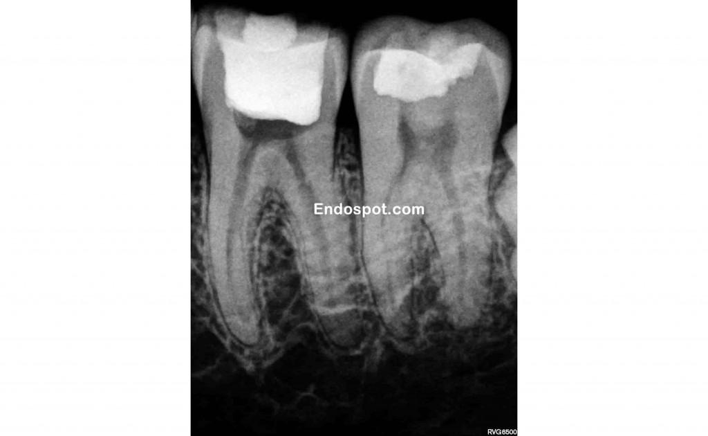
Poor endodontic access

Endodontic access has been performed which gives good access to the canal orifices. But imagine the thickness of dentine that will remain mesially and distally if a crown was prepared for this tooth. The endodontics can be perfectly performed, but the tooth is compromised due to the excessive loss of tooth structure during access.

This is why we aim to keep access limited and you will sometimes see what appears to be incomplete opening of the pulp chamber. This is by design and allows important tooth structure to be maintained that contributes to the strength and durability of the tooth. Strategies for conserving tooth structure during endodontic access include removing restorations and caries and utilising this space for endodontic access. A good discussion of these strategies, along with examples can be found in articles by Clark and Khademi. (3) (4)
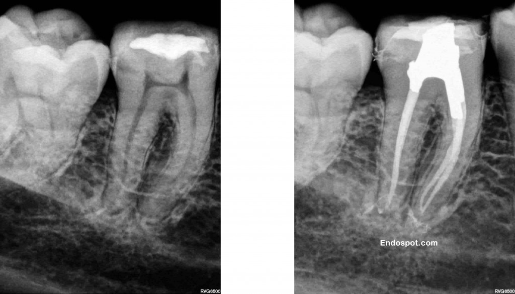
Conservative endodontic access in lower molar

Maintaining the dentine in the peri-cervical region whilst achieving the goals of endodontics give this tooth the best possible chance of survival. Ultrasonics and copious irrigation allow the cleaning of the pulp chamber space and canal spaces without the need for excessive removal of tooth structure.

Maintaining dentine for endodontic access

The restricted access still give good straight line access to the canal orifices. But see how much solid tooth structure remains? It does, however make the job of performing high quality endodontics more difficult. But what’s more important? Us having an easy time of it, or giving the patient the best possible long term outcome?

Limiting tooth destruction becomes a greater challenge when attempting to treat teeth with calcified pulp chambers or canals. When attempting to work through pulp chamber calcification or locate these difficult to find canals it can be easy to remove vitally important dentine.  Assessing the degree of calcification prior to attempting treatment is the key to preventing this iatrogenic damage to teeth. Additionally, if upon access the location of all canals is difficult, it may be better to consider referring at that early stage, rather than to damage the tooth’s long term prognosis in an attempt to locate a difficult to find and prepare canal.

One important aspect of learning to limit the removal of healthy tooth structure for access is that it actually makes endodontic procedures much more difficult (especially in second and third molars) and increases the chances of missing canals, so you need to balance these potential issues with benefits of doing so.

1.    Salehrabi R, Rotstein I. Endodontic treatment outcomes in a large patient population in the USA: An epidemiological study. J Endod. 2004;30(12):846-50.
2.    Vire D. Failure of endodontically treated teeth: classification and evaluation. J Endod. 1991;17:338-42.
3.    Clark D, Khademi J. Modern molar endodontic access and directed dentin conservation. Dent Clin North Am. 2010 Apr;54(2):249-73.
4.    Clark D, Khademi JA. Case studies in modern molar endodontic access and directed dentin conservation. Dent Clin North Am. 2010 Apr;54(2):275-89.